![]()
Products


We are actively taking measures to improve product quality levels.
Applications


Why Hamamatsu?
Resources


Support


Our company


Investors


United Kingdom (EN)
Select your region or country.
High-performance multiphoton excitation microscope using SLM
Observation of deep region of the biological sample by two-photon excitation microscopy
Biological microscopes are used to observe reflections on the surfaces of biological specimens and to observe the transmission of thin sliced biological specimens. Objective lenses of the microscope are designed to have the best condensing condition and best optical performance in these observations. Also, when observing cells, the organelles inside the cell are close to or nearly colorless to the same color, so in many cases, a fluorescence method is used in which only the subject to be observed is stained with a dye and the fluorescence emitted by it is observed.
Recently, in the field of medical and biological sciences, it is required to directly observe deep parts of biological samples. However, if the observation of the deep position of a thick tissue section using biological microscopy is performed, the quality of the condensation by the objective lens will be reduced by deviating from the above design conditions and by generating optical distortion in the observation object itself. This causes blurring in the acquired fluorescent image, which greatly reduces resolution and contrast.
The two-photon excitation fluorescence microscope, which emerged in 1990, is an epoch-making device that can observe the deep part of a biological sample. This microscope uses the phenomenon of two-photon excitation for the emission of fluorescence. Compared with fluorescence observation using confocal fluorescence microscope, in fluorescence observation by two-photon excitation, ultrashort pulsed light of near infrared with high bio-transparency is used for excitation light used for emission of fluorescence, so observation is possible up to the deep part of biological sample. It also has a high depth resolution because the excitation light emits only near the focal point, which is narrowed down by the objective lens. These features are beneficial for observing deep living biological samples.
Three decades have passed since its appearance, and a variety of peripheral technologies have been in place, such as increasing the output and stability of laser light sources that contribute to higher imaging quality, and developing techniques to make living organisms transparent, which enable us to observe deeper sites. Two-photon excitation fluorescence microscopy has begun to be widely used to elucidate biological functions.
Two-photon excitation fluorescence microscope challenges
However, two-photon excitation fluorescence microscopy also has the following challenges:
(1) Image quality deteriorates due to aberrations generated during deep observation of living biological samples.
(2) Long measurement time due to point scan measurement.
(3) Lower resolution than confocal laser microscopy using visible light lasers.
We aim to contribute to medical and biological research by solving these problems using a spatial light modulator (SLM) and realizing high-precision deep observation.
Initiatives to solve problems using SLM
(1) Problem of deteriorating image quality due to aberrations generated when observing the depth of a biological sample
In two-photon excitation fluorescence microscopy, ultrashort pulsed light is made smaller by an objective lens, and light energy is concentrated in a high density in time and space to emit fluorescence. In the observation of the deep part of the biological sample, optical distortion is generated by deviating from the design condition of the objective lens and by being affected by the refractive index mismatch of the light on the surface and inside of the biological sample, and the light energy density is lowered by the blurring of the focal point. Therefore, the spatial resolution is reduced, and at the same time, the brightness of the fluorescence is also reduced, which greatly reduces the quality of the image. Such optical distortion is called aberration.
This aberration must be corrected for observation to obtain a high-resolution image even at the depth of the biological sample. When the SLM is used, the wavefront of the excitation light can be actively controlled to concentrate light in the deep part of the biological sample without the effect of aberration. The important thing to do at this time is to know what aberrations are occurring. This is because aberration is affected by the refractive index difference at the refractive index interface, but it is also affected by the shape of the refractive index interface. We have so far developed a method for correcting aberrations caused by refractive index differences and for correcting aberrations generated by the surface shape of the sample and have confirmed that it is also used for biological samples (Fig. 1).
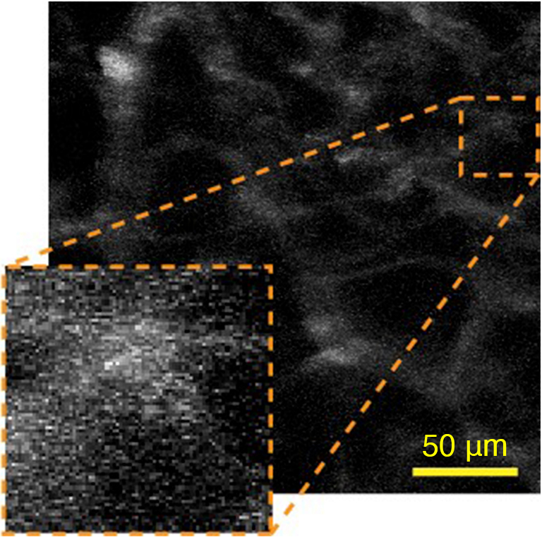
Fig 1. Blood vessel image of transparent mouse brain (depth 1750 µm to 1900 µm)
If correction is made by considering aberrations due to the shape of the biological sample, blood vessels image became clearer (Reference 2).
(a)Applying the aberration correction method we have developed
(b)No aberration correction
(2) Problems with long measurement times
When observing a high-speed phenomenon of a biological sample, the shorter the measurement time, the more advantageous it is. Two-photon excitation fluorescence microscopy uses a galvo scanner, etc., and the system which scans the laser beam and acquires the image is the mainstream. In this scanning imaging, the measurement time is longer compared to the microscope of image illumination. To reduce the measurement time, we have adopted a multi-point simultaneous scanning imaging system, in which the excitation light is branched into multiple points for simultaneous imaging. This multi-point simultaneous scanning imaging was realized by making multiple focal points by controlling the wavefront of the excitation light using the SLM, and by combined it with a multiple location simultaneous detection device, such as a multi-anode photomultiplier tube. We have confirmed that the measurement time can be reduced to one-fourth by branching the excitation light to four points, that there is no effect on the imaging quality, and that deep observation is possible in combination with aberration correction (Fig. 2).
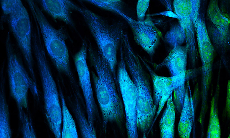
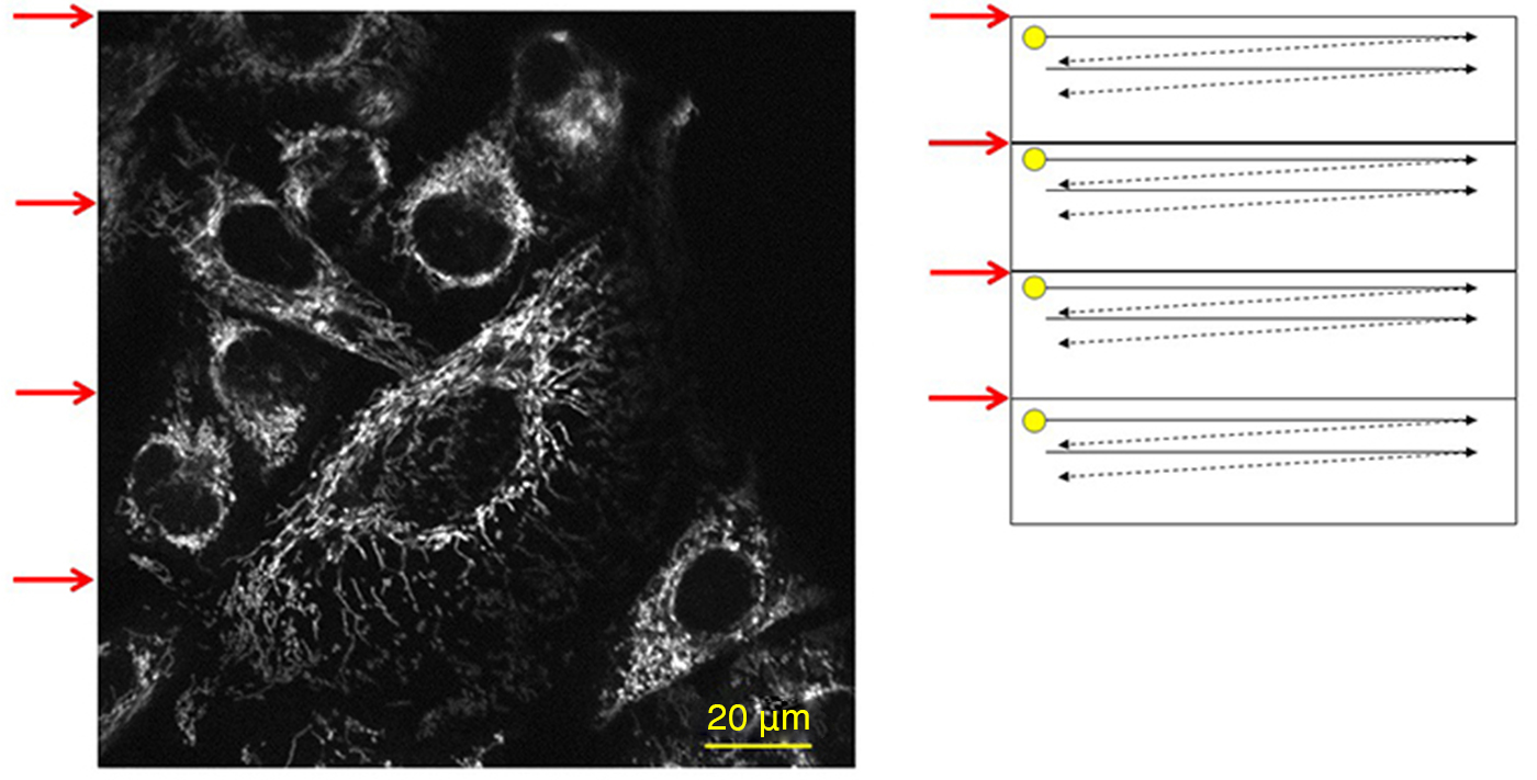
Fig. 2. Fluorescent images obtained by observing Hela cells staining mitochondria with Rhodamine 123
The measurement time has been reduced to 1/4 by performing simultaneous scanning by dividing the excitation light into four sections with SLM.
(3) Lower resolution problem than confocal microscopy using visible light
The resolution of the microscope is higher as the wavelength of the light used is shorter. Two-photon excitation fluorescence microscopy has the advantage of being highly biotransparent and reaching deep region because the wavelength of the excitation light is in the near-infrared region. However, because of its long excitation wavelength, it has a problem of lower resolution compared to confocal microscopy, which is visible light excitation. We are working on ways to increase resolution while maintaining the superiority of deep observations possessed by two-photon excitation fluorescence microscopy. To date, we have developed a method for improving resolution by controlling the intensity distribution of excitation light with the SLM.
Future targets
We are currently working with Hamamatsu University of Medicine on the study of two-photon excitation fluorescence microscopy using the SLM. To date, we have developed aberration corrections suitable for the optical clearing method and conducted observations on the kidney (Fig. 3).
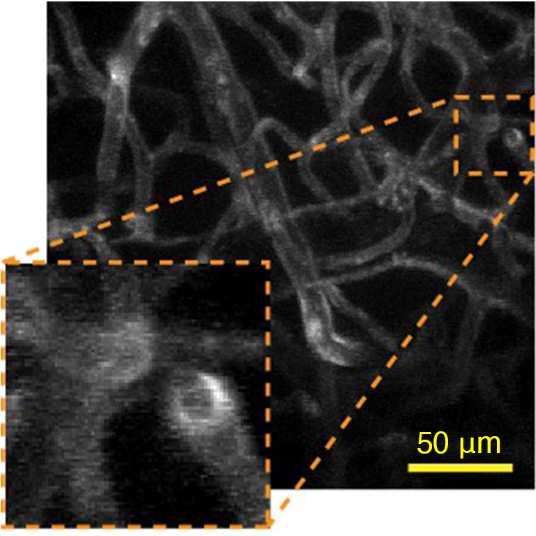
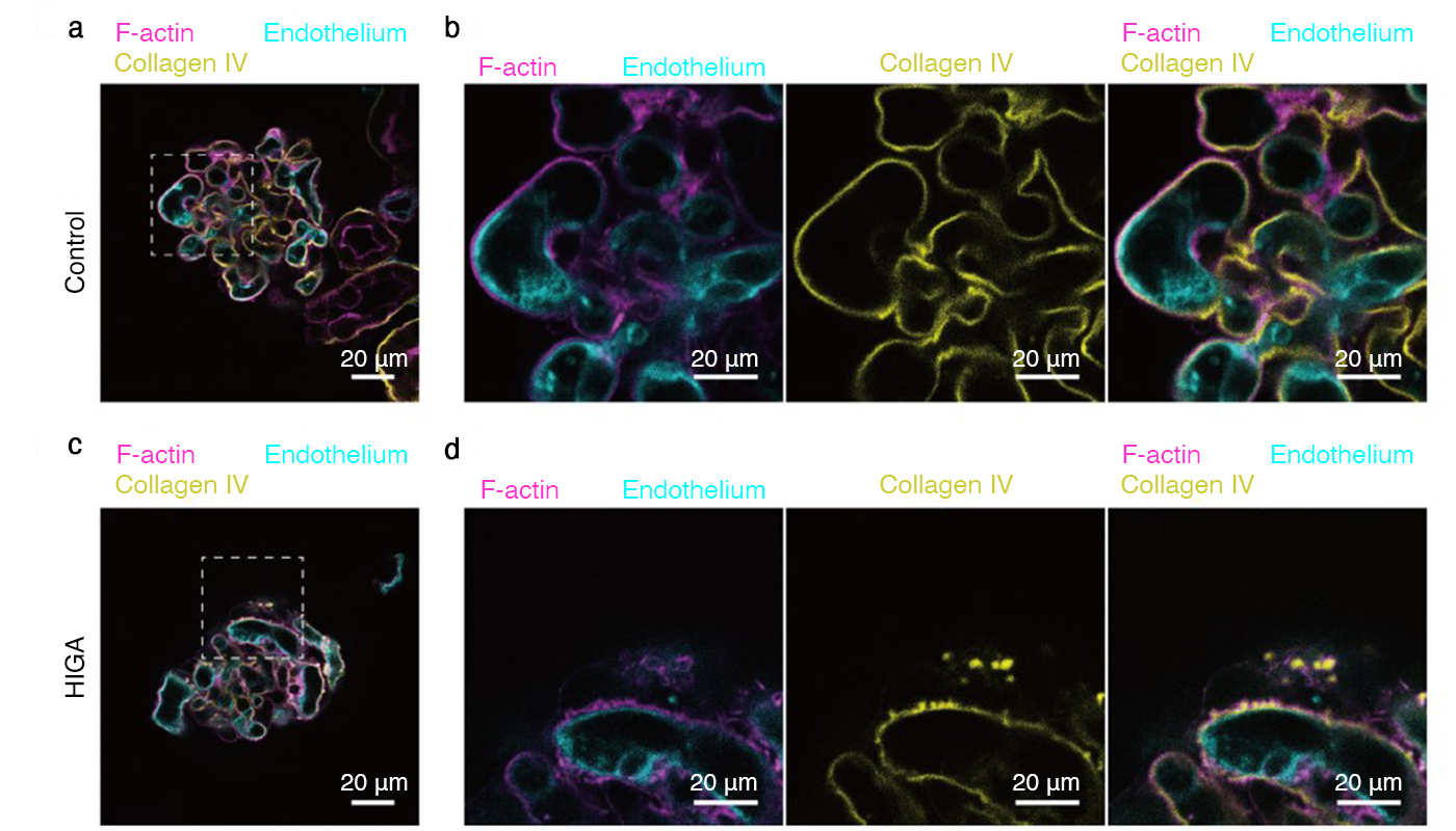
■Images of wild-type (control) and condition-model mouth (HIGA) glomerulus
Renal instrumentation revealed that the protrusions on the vessels in the glomerulus, which were previously confirmed by observation using a conventional electron microscope, were derived from the basement membrane by using a two-photon excitation microscope equipped with the SLM (Reference 5).
■Three-dimensional observation: Mouse renal glomeruli observation cleared by an optical clearing agent
The fixed kidney of mouse was stained, followed by a tissue clearing process and three-dimensional observation. Podocytes* (cells that cover glomeruli) was stained green, blood vessels red, and nuclei blue. In the video, the glomerulus is observed from the surface to the depths while moving the objective lens up and down. By correcting the aberrations caused by the optical clearing agent using an SLM, it is possible to observe them deep even with a commercially available water-immersion objective.
Traditionally, we had made several preparates of kidney by slicing them and viewed them with a light microscope, but slicing had been associated with problems such as deterioration of cross-sectional shape and misalignment. By using the two-photon microscope incorporated with the SLM, it is possible to observe the stereological tissues well, which will make it easier to understand the morphological changes caused by the pathological conditions such as podocyte disorder(Sample was provided by Dr. Alu Konno Department of Microbiology and Immunology, Hamamatsu University School of Medicine).
*Podocytes are cells that cover the periphery of the glomerulus and regulate filtration.
Two-photon excitation fluorescence microscopy will be used more widely in the future to elucidate biofunctions. The SLM of Hamamatsu Photonics is capable of making microscopes multifunctional and high-performance. We aim to contribute to medical and biological research by improving the wavefront control and device technologies, which are peripheral technologies surrounding the microscope and the SLM, so that phenomena that could not be observed before can be discovered.
Reference
- N. Matsumoto, T. Inoue, A. Matsumoto, and S. Okazaki, “Correction of depth-induced spherical aberration for deep observation using two-photon excitation fluorescence microscopy with spatial light modulator,” Biomed. Opt. Express 6(7) 2575-2587 (2015).
- N. Matsumoto, A. Konno, T. Inoue and S. Okazaki, “Aberration correction considering curved sample surface shape for non-contact two-photon excitation microscopy with spatial light modulator,” Sci. Rep. 9252 (2018).
- N. Matsumoto, A. Konno, Y. Ohbayashi, T. Inoue, A. Matsumoto, K. Uchimura, K. Kadomatsu, and S. Okazaki, “Correction of spherical aberration in multi-focal multiphoton microscopy with spatial light modulator,” 25(6), 7055-7068 (2017).
- N. Matsumoto, A. Konno, T. Inoue, K. Watanabe, and S. Okazaki, “Amplitude-modulation-type multi-ring mask for two-photon excitation scanning microscopy,” OSA continuum 4(6), 1696-1711 (2021).
- A. Konno, N. Matsumoto, Y. Tomono, and S. Okazaki, “Pathological application of carbocyanine dye-based multicolour imaging of vasculature and associated structures,” Sci. Rep. 10, 12613 (2020).
- Confirmation
-
It looks like you're in the . If this is not your location, please select the correct region or country below.
You're headed to Hamamatsu Photonics website for GB (English). If you want to view an other country's site, the optimized information will be provided by selecting options below.
In order to use this website comfortably, we use cookies. For cookie details please see our cookie policy.
- Cookie Policy
-
This website or its third-party tools use cookies, which are necessary to its functioning and required to achieve the purposes illustrated in this cookie policy. By closing the cookie warning banner, scrolling the page, clicking a link or continuing to browse otherwise, you agree to the use of cookies.
Hamamatsu uses cookies in order to enhance your experience on our website and ensure that our website functions.
You can visit this page at any time to learn more about cookies, get the most up to date information on how we use cookies and manage your cookie settings. We will not use cookies for any purpose other than the ones stated, but please note that we reserve the right to update our cookies.
1. What are cookies?
For modern websites to work according to visitor’s expectations, they need to collect certain basic information about visitors. To do this, a site will create small text files which are placed on visitor’s devices (computer or mobile) - these files are known as cookies when you access a website. Cookies are used in order to make websites function and work efficiently. Cookies are uniquely assigned to each visitor and can only be read by a web server in the domain that issued the cookie to the visitor. Cookies cannot be used to run programs or deliver viruses to a visitor’s device.
Cookies do various jobs which make the visitor’s experience of the internet much smoother and more interactive. For instance, cookies are used to remember the visitor’s preferences on sites they visit often, to remember language preference and to help navigate between pages more efficiently. Much, though not all, of the data collected is anonymous, though some of it is designed to detect browsing patterns and approximate geographical location to improve the visitor experience.
Certain type of cookies may require the data subject’s consent before storing them on the computer.
2. What are the different types of cookies?
This website uses two types of cookies:
- First party cookies. For our website, the first party cookies are controlled and maintained by Hamamatsu. No other parties have access to these cookies.
- Third party cookies. These cookies are implemented by organizations outside Hamamatsu. We do not have access to the data in these cookies, but we use these cookies to improve the overall website experience.
3. How do we use cookies?
This website uses cookies for following purposes:
- Certain cookies are necessary for our website to function. These are strictly necessary cookies and are required to enable website access, support navigation or provide relevant content. These cookies direct you to the correct region or country, and support security and ecommerce. Strictly necessary cookies also enforce your privacy preferences. Without these strictly necessary cookies, much of our website will not function.
- Analytics cookies are used to track website usage. This data enables us to improve our website usability, performance and website administration. In our analytics cookies, we do not store any personal identifying information.
- Functionality cookies. These are used to recognize you when you return to our website. This enables us to personalize our content for you, greet you by name and remember your preferences (for example, your choice of language or region).
- These cookies record your visit to our website, the pages you have visited and the links you have followed. We will use this information to make our website and the advertising displayed on it more relevant to your interests. We may also share this information with third parties for this purpose.
Cookies help us help you. Through the use of cookies, we learn what is important to our visitors and we develop and enhance website content and functionality to support your experience. Much of our website can be accessed if cookies are disabled, however certain website functions may not work. And, we believe your current and future visits will be enhanced if cookies are enabled.
4. Which cookies do we use?
There are two ways to manage cookie preferences.
- You can set your cookie preferences on your device or in your browser.
- You can set your cookie preferences at the website level.
If you don’t want to receive cookies, you can modify your browser so that it notifies you when cookies are sent to it or you can refuse cookies altogether. You can also delete cookies that have already been set.
If you wish to restrict or block web browser cookies which are set on your device then you can do this through your browser settings; the Help function within your browser should tell you how. Alternatively, you may wish to visit www.aboutcookies.org, which contains comprehensive information on how to do this on a wide variety of desktop browsers.
5. What are Internet tags and how do we use them with cookies?
Occasionally, we may use internet tags (also known as action tags, single-pixel GIFs, clear GIFs, invisible GIFs and 1-by-1 GIFs) at this site and may deploy these tags/cookies through a third-party advertising partner or a web analytical service partner which may be located and store the respective information (including your IP-address) in a foreign country. These tags/cookies are placed on both online advertisements that bring users to this site and on different pages of this site. We use this technology to measure the visitors' responses to our sites and the effectiveness of our advertising campaigns (including how many times a page is opened and which information is consulted) as well as to evaluate your use of this website. The third-party partner or the web analytical service partner may be able to collect data about visitors to our and other sites because of these internet tags/cookies, may compose reports regarding the website’s activity for us and may provide further services which are related to the use of the website and the internet. They may provide such information to other parties if there is a legal requirement that they do so, or if they hire the other parties to process information on their behalf.
If you would like more information about web tags and cookies associated with on-line advertising or to opt-out of third-party collection of this information, please visit the Network Advertising Initiative website http://www.networkadvertising.org.
6. Analytics and Advertisement Cookies
We use third-party cookies (such as Google Analytics) to track visitors on our website, to get reports about how visitors use the website and to inform, optimize and serve ads based on someone's past visits to our website.
You may opt-out of Google Analytics cookies by the websites provided by Google:
https://tools.google.com/dlpage/gaoptout?hl=en
As provided in this Privacy Policy (Article 5), you can learn more about opt-out cookies by the website provided by Network Advertising Initiative:
http://www.networkadvertising.org
We inform you that in such case you will not be able to wholly use all functions of our website.
Close