![]()
Products


We are actively taking measures to improve product quality levels.
Applications


Why Hamamatsu?
Resources


Support


Our company


Investors


United States (EN)
Select your region or country.


Dental imaging
Dental is one of the largest markets for X-ray imaging, and Hamamatsu has the widest range of dental X-ray sensor options available today. You can learn about dental applications and our product offerings by exploring the pages and links below.
First dental radiograph (1896)
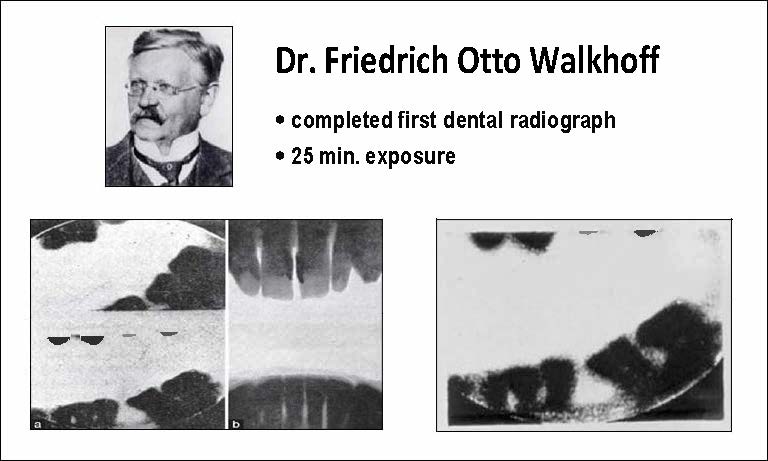
*Dr. Otto Walkhoff, German dentist, and the first dental radiograph, an image of his own teeth (1896). Photo courtesy of Medcrave.
Before the dawn of the 20th century, on December 22, 1895, William C. Roentgen discovered X-rays and took the first X-ray image ever, an image of his wife’s hand. However, within just two weeks, Otto Walkhoff, a German dentist, captured the first radiograph of his own teeth. Walkhoff generated the first intraoral radiograph, after a 25-minute exposure. In his account, he reported: “It was a real torture but I felt tremendously happy when I saw the results, and weighed the importance of Roentgen’s discovery on the future of dentistry.”
Since then, dental radiography has become the standard of care for diagnosing disease and other health issues associated with teeth and the mouth in general.
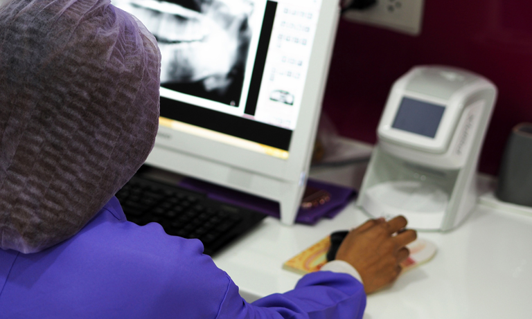
Dental X-ray imaging technique has evolved over the years and is currently defined as standard radiography, panoramic imaging or cephalometric imaging. Standard radiography started with film, but in recent years more advanced imaging techniques have been developed: first with computed radiography (CR) systems and, more recently, with digital radiography (DR) sensors.
Furthermore, two specialized applications of dental radiography have emerged. Panoramic imaging, though not considered diagnostic quality, allows the dentist to observe all of the patient’s teeth in one large frontal-view image to do a quick screening. Cephalometric imaging is another radiographic technique; however, these images are taken from the side of a patient’s head and used mostly by orthodontists during treatment planning.
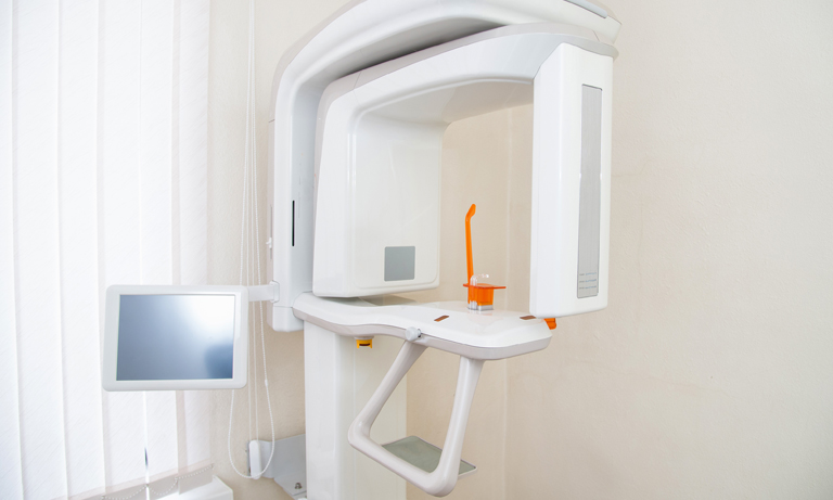
In addition, 3-dimensional imaging of teeth has been achieved by adapting cone beam computed tomography (CBCT) technology for dental use. CBCT systems offer dentists greater clarity and detail of latent dental health issues, as well as providing a valuable tool for planning implant and other dental surgeries.
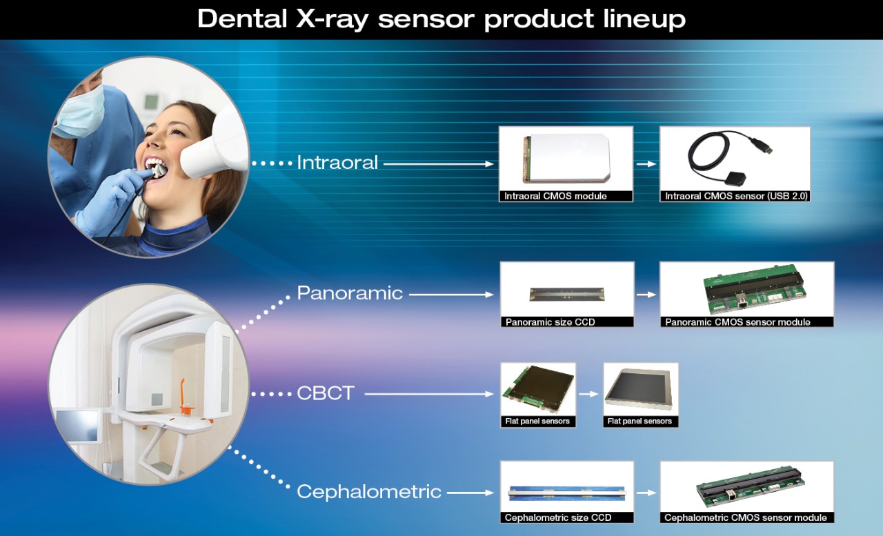
A wide range of image sensors for dental X-ray imaging
Overall, Hamamatsu has the widest and most advanced selection of image sensors for dental X-ray imaging available today. We work closely with engineers and entrepreneurs to adopt our products and deliver the most advanced digital dental X-ray imaging systems available today. You can learn more about each imaging technique below and see how Hamamatsu’s line of advanced, ultra-reliable sensors can improve the quality and performance of your dental X-ray imaging system.
Note: We only accept inquiries from OEMS and System Integrators who can manage regulatory compliance for the medical device they have, that would integrate our sensors.
Hamamatsu is the only company in the world providing digital sensors for every X-ray dental imaging modality
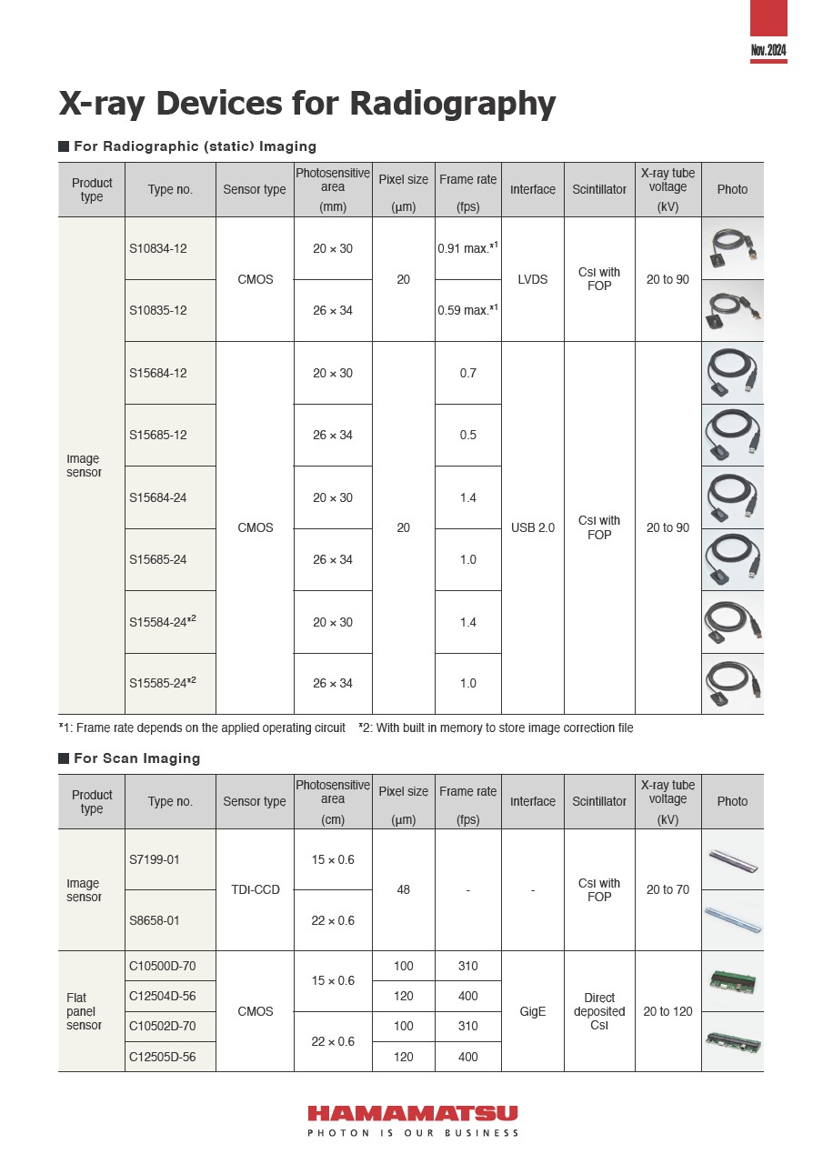
Hamamatsu has data sheets on all products listed on these pages. We also offer this convenient selection guide of dental X-ray sensors. If you are considering to design a new OEM dental X-ray imaging system, please email us or contact your local Hamamatsu office to request a copy.
Disclaimer: The products listed on this website are not finished medical devices and do not have clearance from the FDA or any other regulating body. These products are only offered as components in OEM X-ray imaging systems, and it is incumbent on the OEM to obtain 510K clearance for any medical diagnostic use. These products are not intended for sale to dentists or other healthcare practitioners.
Intraoral computed radiography (CR)
Considered to be a bridge between classical radiography and increasingly popular fully digital methods, computed radiography offers the comfort of X-ray film with the benefit of high quality digitized X-ray images.
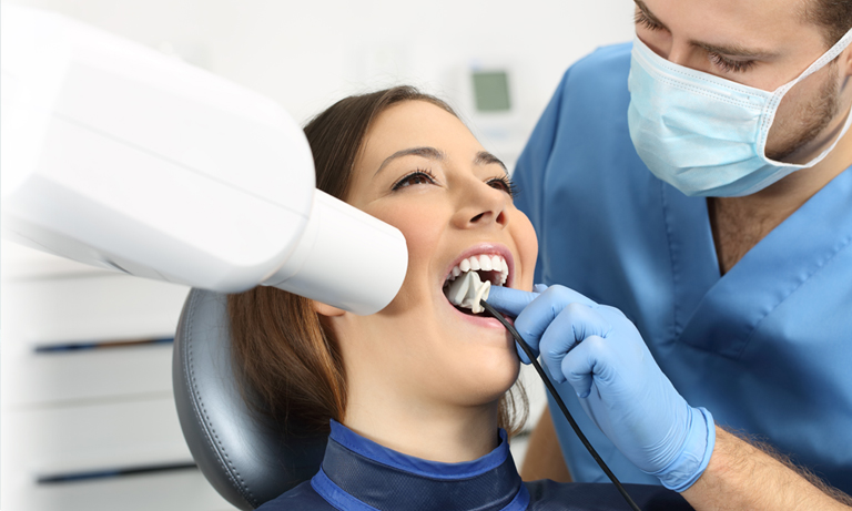
Intraoral digital radiography
Digital radiography offers high quality imaging with the convenience of a simple-to-use, compact X-ray sensor.
Panoramic and cephalometric imaging
Panoramic and cephalometric imaging offer the convenience of seeing the entire mouth in one digital X-ray image from different perspectives. Panoramic imaging provides a frontal view while cephalometric imaging provides a profile view of the mouth and teeth.
Cone beam computed tomography (CBCT)
Cone beam computed tomography (CBCT) is a modern imaging technique adopted by the dental industry, providing doctors and dentists with advanced diagnostic and surgical planning tools.

Custom intraoral sensors
Hamamatsu offers custom, high resolution CMOS sensors for dental and veterinary intraoral use. We offer a wide range of customization capabilities.
Research and development
In order to contribute to a healthy society, we apply a wide range of photonics technologies into healthcare and medicine. Our Central Research Laboratory continually researches and develops the best components for PET, near-infrared spectroscopy and imaging, sports measurements, and biomedical research. This work will continue to expand the new possibilities of light into the future. Learn more about initiatives from our Central Research Laboratory.

Are you a dental OEM with an idea for a custom intraoral image sensor? Hamamatsu offers a wide range of advanced features and options.
- Confirmation
-
It looks like you're in the . If this is not your location, please select the correct region or country below.
You're headed to Hamamatsu Photonics website for US (English). If you want to view an other country's site, the optimized information will be provided by selecting options below.
In order to use this website comfortably, we use cookies. For cookie details please see our cookie policy.
- Cookie Policy
-
This website or its third-party tools use cookies, which are necessary to its functioning and required to achieve the purposes illustrated in this cookie policy. By closing the cookie warning banner, scrolling the page, clicking a link or continuing to browse otherwise, you agree to the use of cookies.
Hamamatsu uses cookies in order to enhance your experience on our website and ensure that our website functions.
You can visit this page at any time to learn more about cookies, get the most up to date information on how we use cookies and manage your cookie settings. We will not use cookies for any purpose other than the ones stated, but please note that we reserve the right to update our cookies.
1. What are cookies?
For modern websites to work according to visitor’s expectations, they need to collect certain basic information about visitors. To do this, a site will create small text files which are placed on visitor’s devices (computer or mobile) - these files are known as cookies when you access a website. Cookies are used in order to make websites function and work efficiently. Cookies are uniquely assigned to each visitor and can only be read by a web server in the domain that issued the cookie to the visitor. Cookies cannot be used to run programs or deliver viruses to a visitor’s device.
Cookies do various jobs which make the visitor’s experience of the internet much smoother and more interactive. For instance, cookies are used to remember the visitor’s preferences on sites they visit often, to remember language preference and to help navigate between pages more efficiently. Much, though not all, of the data collected is anonymous, though some of it is designed to detect browsing patterns and approximate geographical location to improve the visitor experience.
Certain type of cookies may require the data subject’s consent before storing them on the computer.
2. What are the different types of cookies?
This website uses two types of cookies:
- First party cookies. For our website, the first party cookies are controlled and maintained by Hamamatsu. No other parties have access to these cookies.
- Third party cookies. These cookies are implemented by organizations outside Hamamatsu. We do not have access to the data in these cookies, but we use these cookies to improve the overall website experience.
3. How do we use cookies?
This website uses cookies for following purposes:
- Certain cookies are necessary for our website to function. These are strictly necessary cookies and are required to enable website access, support navigation or provide relevant content. These cookies direct you to the correct region or country, and support security and ecommerce. Strictly necessary cookies also enforce your privacy preferences. Without these strictly necessary cookies, much of our website will not function.
- Analytics cookies are used to track website usage. This data enables us to improve our website usability, performance and website administration. In our analytics cookies, we do not store any personal identifying information.
- Functionality cookies. These are used to recognize you when you return to our website. This enables us to personalize our content for you, greet you by name and remember your preferences (for example, your choice of language or region).
- These cookies record your visit to our website, the pages you have visited and the links you have followed. We will use this information to make our website and the advertising displayed on it more relevant to your interests. We may also share this information with third parties for this purpose.
Cookies help us help you. Through the use of cookies, we learn what is important to our visitors and we develop and enhance website content and functionality to support your experience. Much of our website can be accessed if cookies are disabled, however certain website functions may not work. And, we believe your current and future visits will be enhanced if cookies are enabled.
4. Which cookies do we use?
There are two ways to manage cookie preferences.
- You can set your cookie preferences on your device or in your browser.
- You can set your cookie preferences at the website level.
If you don’t want to receive cookies, you can modify your browser so that it notifies you when cookies are sent to it or you can refuse cookies altogether. You can also delete cookies that have already been set.
If you wish to restrict or block web browser cookies which are set on your device then you can do this through your browser settings; the Help function within your browser should tell you how. Alternatively, you may wish to visit www.aboutcookies.org, which contains comprehensive information on how to do this on a wide variety of desktop browsers.
5. What are Internet tags and how do we use them with cookies?
Occasionally, we may use internet tags (also known as action tags, single-pixel GIFs, clear GIFs, invisible GIFs and 1-by-1 GIFs) at this site and may deploy these tags/cookies through a third-party advertising partner or a web analytical service partner which may be located and store the respective information (including your IP-address) in a foreign country. These tags/cookies are placed on both online advertisements that bring users to this site and on different pages of this site. We use this technology to measure the visitors' responses to our sites and the effectiveness of our advertising campaigns (including how many times a page is opened and which information is consulted) as well as to evaluate your use of this website. The third-party partner or the web analytical service partner may be able to collect data about visitors to our and other sites because of these internet tags/cookies, may compose reports regarding the website’s activity for us and may provide further services which are related to the use of the website and the internet. They may provide such information to other parties if there is a legal requirement that they do so, or if they hire the other parties to process information on their behalf.
If you would like more information about web tags and cookies associated with on-line advertising or to opt-out of third-party collection of this information, please visit the Network Advertising Initiative website http://www.networkadvertising.org.
6. Analytics and Advertisement Cookies
We use third-party cookies (such as Google Analytics) to track visitors on our website, to get reports about how visitors use the website and to inform, optimize and serve ads based on someone's past visits to our website.
You may opt-out of Google Analytics cookies by the websites provided by Google:
https://tools.google.com/dlpage/gaoptout?hl=en
As provided in this Privacy Policy (Article 5), you can learn more about opt-out cookies by the website provided by Network Advertising Initiative:
http://www.networkadvertising.org
We inform you that in such case you will not be able to wholly use all functions of our website.
Close