![]()
Products


We are actively taking measures to improve product quality levels.
Applications


Why Hamamatsu?
Resources


Support


Our company


Investors


United States (EN)
Select your region or country.
High temporal performance gamma-ray detector
Detector concept
Cherenkov emission has received attention to improve timing resolution of detectors dedicated to positron emission tomography. Since the Cherenkov photons are promptly emitted within an order of 1 ps, the timing performance of the detector would be enhanced compared to a scintillation-based detector. A monolithic Cherenkov radiator which does not emit scintillation photons was employed to suppress reflections in the radiator. However, it is challenging to reconstruct the interaction position of the gamma ray because the number of Cherenkov photons emitted when a gamma ray interacts with the radiator is only 30 at most. As a photodetector, a high-dense photodetector array with individual readout was applied to extract all temporal and spatial information of the Cherenkov photons.

■Features
Monolithic Cherenkov radiator
→To maximize the timing performance of Cherenkov emission
Individual readout
→To obtain all temporal- and spatial-information from a few Cherenkov photons
Required specification for photodetector
Monte Carlo simulations were performed [Publication 1]. According to the simulations, we found that requirements for the photodetector are high-dense and good single photon time resolution (SPTR).For example, the SPTR must be better than σ = 40 ps to obtain a coincidence time resolution (CTR) of 100 ps full width at half maximum (FWHM).
Moreover, we have investigated potential capability of deep learning to improve the position resolution of the proposed detector [Publication 2].
Experimental validation
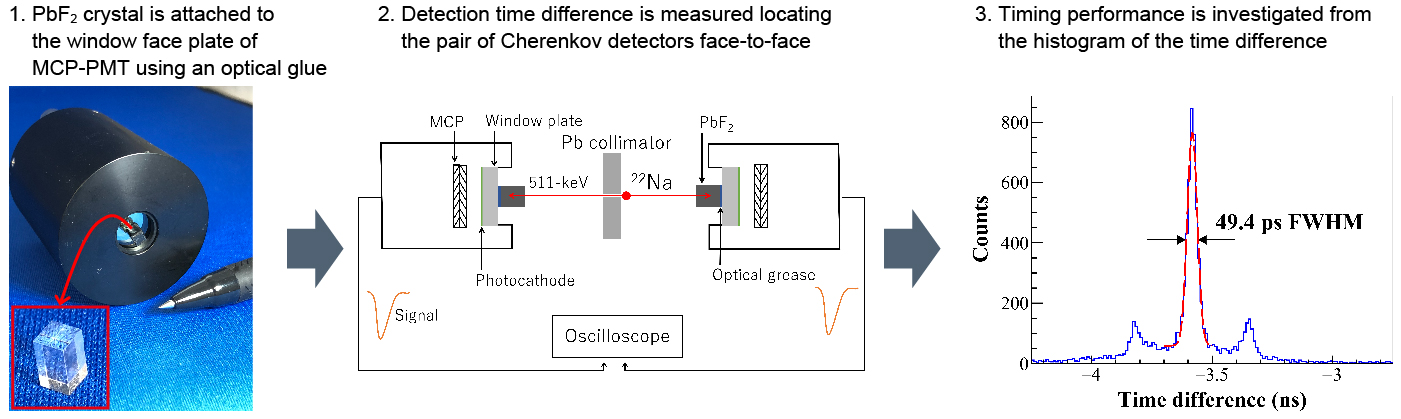
An experiment measuring the CTR was performed using a high speed photodetector of MCP-PMT (R3809). The SPTR of the MCP-PMT is 25 ps FWHM.
A lead fluoride Cherenkov radiator was attached to the window face plate of the MCP-PMT using an optical glue.
As a result, the CTR better than 50 ps FWHM was successfully obtained, and this result is consistent with the simulation result [Publication 3].
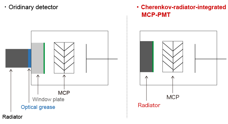
Development of Cherenkov-radiator-integrated MCP-PMT
There are optical boundaries in the Cherenkov detector described above. The optical boundaries will worsen the timing performance of the detector. We have developed a Cherenkov-radiator-integrated MCP-PMT (CRI-MCP-PMT) to remove the optical boundaries from the detector. The window face plate of the ordinary MCP-PMT was replaced with the Cherenkov radiator and Al2O3 layer was inserted between the radiator and the photocathode using atomic layer deposition technique.
The CTR was measured using a pair of the CRI-MCP-PMTs, and the CTR of 41.9 ps FWHM was obtained [Publication 4]. Additionally, the CTR was improved to 30.1 ps FWHM by optimizing the analysis parameters, e.g. timing pick-off threshold level and pulse area.
Performance improvement with lead-free materials
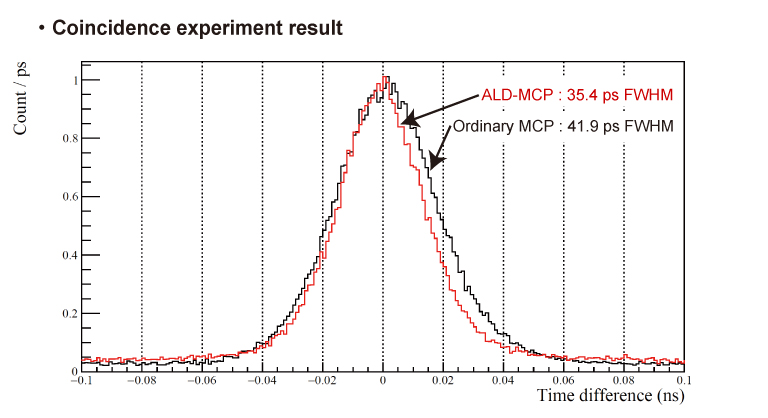
By reviewing all materials starting from scatch and applying an ALD technique, we succeeded in creating ALD-MCPs that ensure low noise and high signal multiplication (gain) without using materials containing lead. Eliminating lead is great step forward because its use is restricted in the RoHS directive issued by the European Union (EU) as a hazardous substance.

A coincidence experiment revealed that a PMT integrating the ALD-MCP can enhance the CTR from 41.9 to 35.4 ps FWHM compared to an ordinary MCP-PMT [reference 6].
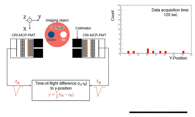
Reconstruction-free imaging
We have succeeded in being the first in the world to achieve high-accuracy positron emission imaging without image reconstruction by utilizing the pair of detectors mentioned above, and unique light detection and signal processing techniques [reference 7]. Applying these successful research results are promising to achieve a completely innovative new type of radiation medical imaging system capable of making speedy diagnoses from a simple, compact setup yet with the same or higher accuracy than currently used radiation imaging systems such as positron emission tomography (PET). This will help boost inspection efficiency for detecting diseased tissues or organs, such as from cancer, and also reduce the radiation exposure dose, alleviating the load on the patient and medical staff. We will further improve the detector performance to make this new concept more practical.
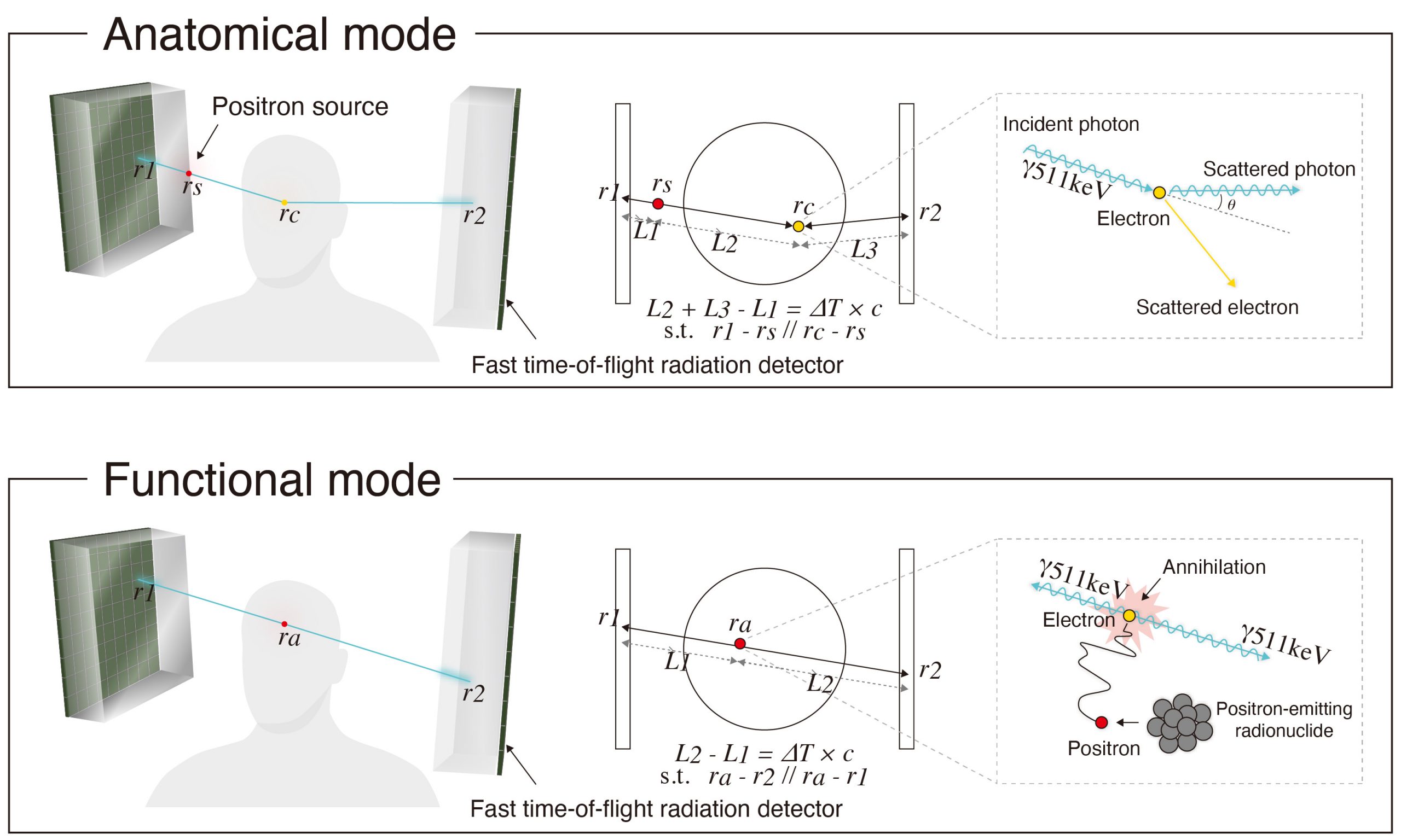
Reconstruction-free anatomical imaging method
PET scanners are generally combined with an X-ray computed tomography (X-CT). The data obtained by a PET scanner contains metabolic information of a patient, however, does not contain its anatomical information, which is necessary to correct the PET data and make it more quantitative. The above-mentioned proposed reconstruction-free imaging method using ultrafast timing detectors also requires the anatomical information because it is also a system measuring patient’s metabolic information. Although the anatomical information can be obtained using X-CTs, X-CTs generally are huge systems, which will not be able to maximize the potential of the compactness of the proposed reconstruction-free imaging method.
To tackle with this problem, we propose a new anatomical imaging method using the same ultrafast timing detectors to maintain the potential [reference 9]. We utilize a nature of an interaction between matter and light, so called Compton scattering. By utilizing the fact that probability of Compton scattering between matter and light is proportional to the electron density of the matter, we devised an algorithm which can localize the three-dimensional interaction position based on spatiotemporal information of the ultrafast detectors. It is demonstrated that the anatomical information can be obtained by our algorithm through a Monte Carlo simulation.
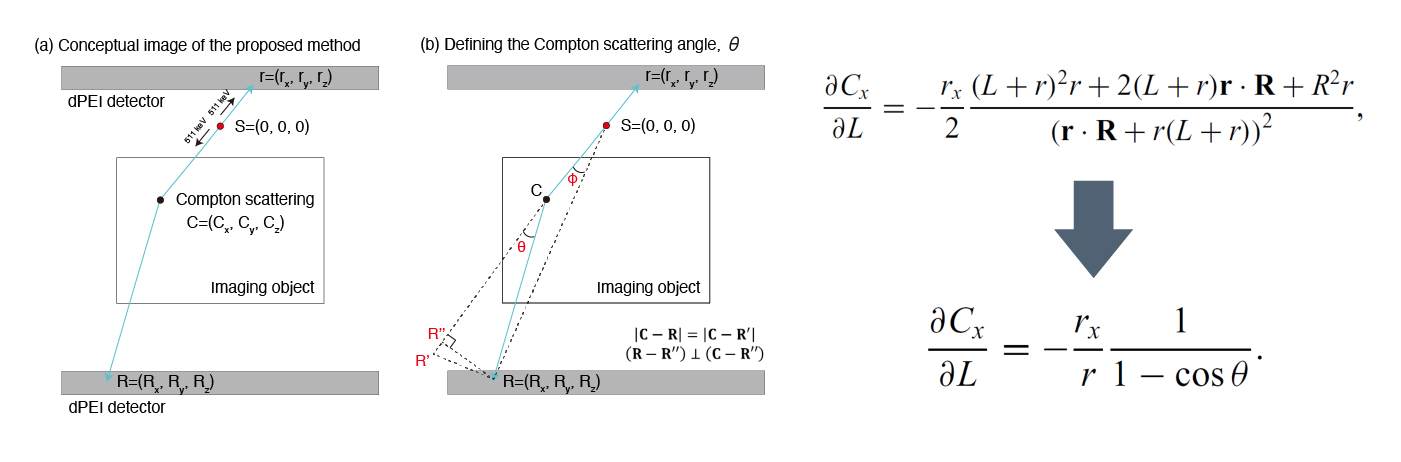
Theoretical understanding of anatomical imaging method
Although it was proved that an anatomical image can be obtained by our proposed algorithm, we also found that the spatial resolution of the anatomical image obtained by our proposed algorithm is significantly worse than that intuitively expected. Therefore, we worked on understanding a physics behind our proposed algorithm [reference 10]. Through the theoretical approach, we figure out that the spatial resolution of the anatomical image gets worse in certain conditions. These conditions are validated through Monte Carlo simulations, and we succeeded in improving the spatial resolution of the anatomical image be eliminating the conditions.
Publications
1. R. Ota et al., Med. Phys. 45 (2018) pp. 1999-2008
2. F. Hashimoto et al., Biomed. Phys. Eng. Express 5 (2019) 035001
3. R. Ota et al., Nucl. Inst. Meth. A 923 (2019) pp. 1-4
4. R. Ota et al., Phys Med. Biol. 64 (2019) 07LT01
5. R. Ota et al., Phys. Med. biol. 65 (2020) 10NT03
6. R. Ota et al., Phys Med. Biol. 66 (2021) 064006
7. S. I. Kwon, R. Ota, et al., Nat. Photon. 15 (2021) 914-918
8. Y. Onishi et al., Phys Med. Biol. 67 (2022) 04NT01
9. Y. Onishi et al., IEEE Trans. Med. Imaging 43 (2024) 1654-1663
10. Y. Onishi and R. Ota, Phys. Rev. Applied 22 (2024) 024049
Patents
US Patent 10,795,035
US Patent 10,816,682
US Patent 10,996,348
US Patent 10,925,557
- Confirmation
-
It looks like you're in the . If this is not your location, please select the correct region or country below.
You're headed to Hamamatsu Photonics website for US (English). If you want to view an other country's site, the optimized information will be provided by selecting options below.
In order to use this website comfortably, we use cookies. For cookie details please see our cookie policy.
- Cookie Policy
-
This website or its third-party tools use cookies, which are necessary to its functioning and required to achieve the purposes illustrated in this cookie policy. By closing the cookie warning banner, scrolling the page, clicking a link or continuing to browse otherwise, you agree to the use of cookies.
Hamamatsu uses cookies in order to enhance your experience on our website and ensure that our website functions.
You can visit this page at any time to learn more about cookies, get the most up to date information on how we use cookies and manage your cookie settings. We will not use cookies for any purpose other than the ones stated, but please note that we reserve the right to update our cookies.
1. What are cookies?
For modern websites to work according to visitor’s expectations, they need to collect certain basic information about visitors. To do this, a site will create small text files which are placed on visitor’s devices (computer or mobile) - these files are known as cookies when you access a website. Cookies are used in order to make websites function and work efficiently. Cookies are uniquely assigned to each visitor and can only be read by a web server in the domain that issued the cookie to the visitor. Cookies cannot be used to run programs or deliver viruses to a visitor’s device.
Cookies do various jobs which make the visitor’s experience of the internet much smoother and more interactive. For instance, cookies are used to remember the visitor’s preferences on sites they visit often, to remember language preference and to help navigate between pages more efficiently. Much, though not all, of the data collected is anonymous, though some of it is designed to detect browsing patterns and approximate geographical location to improve the visitor experience.
Certain type of cookies may require the data subject’s consent before storing them on the computer.
2. What are the different types of cookies?
This website uses two types of cookies:
- First party cookies. For our website, the first party cookies are controlled and maintained by Hamamatsu. No other parties have access to these cookies.
- Third party cookies. These cookies are implemented by organizations outside Hamamatsu. We do not have access to the data in these cookies, but we use these cookies to improve the overall website experience.
3. How do we use cookies?
This website uses cookies for following purposes:
- Certain cookies are necessary for our website to function. These are strictly necessary cookies and are required to enable website access, support navigation or provide relevant content. These cookies direct you to the correct region or country, and support security and ecommerce. Strictly necessary cookies also enforce your privacy preferences. Without these strictly necessary cookies, much of our website will not function.
- Analytics cookies are used to track website usage. This data enables us to improve our website usability, performance and website administration. In our analytics cookies, we do not store any personal identifying information.
- Functionality cookies. These are used to recognize you when you return to our website. This enables us to personalize our content for you, greet you by name and remember your preferences (for example, your choice of language or region).
- These cookies record your visit to our website, the pages you have visited and the links you have followed. We will use this information to make our website and the advertising displayed on it more relevant to your interests. We may also share this information with third parties for this purpose.
Cookies help us help you. Through the use of cookies, we learn what is important to our visitors and we develop and enhance website content and functionality to support your experience. Much of our website can be accessed if cookies are disabled, however certain website functions may not work. And, we believe your current and future visits will be enhanced if cookies are enabled.
4. Which cookies do we use?
There are two ways to manage cookie preferences.
- You can set your cookie preferences on your device or in your browser.
- You can set your cookie preferences at the website level.
If you don’t want to receive cookies, you can modify your browser so that it notifies you when cookies are sent to it or you can refuse cookies altogether. You can also delete cookies that have already been set.
If you wish to restrict or block web browser cookies which are set on your device then you can do this through your browser settings; the Help function within your browser should tell you how. Alternatively, you may wish to visit www.aboutcookies.org, which contains comprehensive information on how to do this on a wide variety of desktop browsers.
5. What are Internet tags and how do we use them with cookies?
Occasionally, we may use internet tags (also known as action tags, single-pixel GIFs, clear GIFs, invisible GIFs and 1-by-1 GIFs) at this site and may deploy these tags/cookies through a third-party advertising partner or a web analytical service partner which may be located and store the respective information (including your IP-address) in a foreign country. These tags/cookies are placed on both online advertisements that bring users to this site and on different pages of this site. We use this technology to measure the visitors' responses to our sites and the effectiveness of our advertising campaigns (including how many times a page is opened and which information is consulted) as well as to evaluate your use of this website. The third-party partner or the web analytical service partner may be able to collect data about visitors to our and other sites because of these internet tags/cookies, may compose reports regarding the website’s activity for us and may provide further services which are related to the use of the website and the internet. They may provide such information to other parties if there is a legal requirement that they do so, or if they hire the other parties to process information on their behalf.
If you would like more information about web tags and cookies associated with on-line advertising or to opt-out of third-party collection of this information, please visit the Network Advertising Initiative website http://www.networkadvertising.org.
6. Analytics and Advertisement Cookies
We use third-party cookies (such as Google Analytics) to track visitors on our website, to get reports about how visitors use the website and to inform, optimize and serve ads based on someone's past visits to our website.
You may opt-out of Google Analytics cookies by the websites provided by Google:
https://tools.google.com/dlpage/gaoptout?hl=en
As provided in this Privacy Policy (Article 5), you can learn more about opt-out cookies by the website provided by Network Advertising Initiative:
http://www.networkadvertising.org
We inform you that in such case you will not be able to wholly use all functions of our website.
Close