
Products












We are actively taking measures to improve product quality levels.
Applications






Why Hamamatsu?
Resources



Support



Our company





Investors








United States (EN)
Select your region or country.
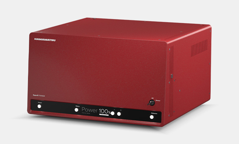
MoxiePlex Multi-spectral imaging system
C16919-01
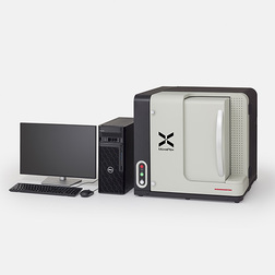
The MoxiePlex Multi-spectral imaging system captures high-resolution images of entire tissue samples stained with multiple fluorescent dyes. These images support spatial proteomics workflows and accelerate translational research in drug discovery.
- Acquire high-resolution images with up to 9 channels
- Imaging examples by reagent product
- Automated sample recognition and exposure time calculation
- Unmixing technology for multiplex fluorescence analysis
- Library creation for enhanced unmixing accuracy
- Related documents
- Related videos
- Specifications
- Dimensions
Acquire high-resolution images with up to 9 channels
The MoxiePlex provides up to 9 channels of fluorescence imaging across the entire tissue section on a glass slide.
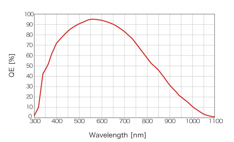
With a 16-bit output, it delivers high-resolution imaging essential for image analysis. Additionally, the system is equipped with a state-of-the-art camera featuring high quantum efficiency across a 400 nm to 900 nm wavelength range, allowing for a broader selection of fluorescent reagents and enhanced biomarker detection.
Spectral sensitivity
Imaging examples by reagent product
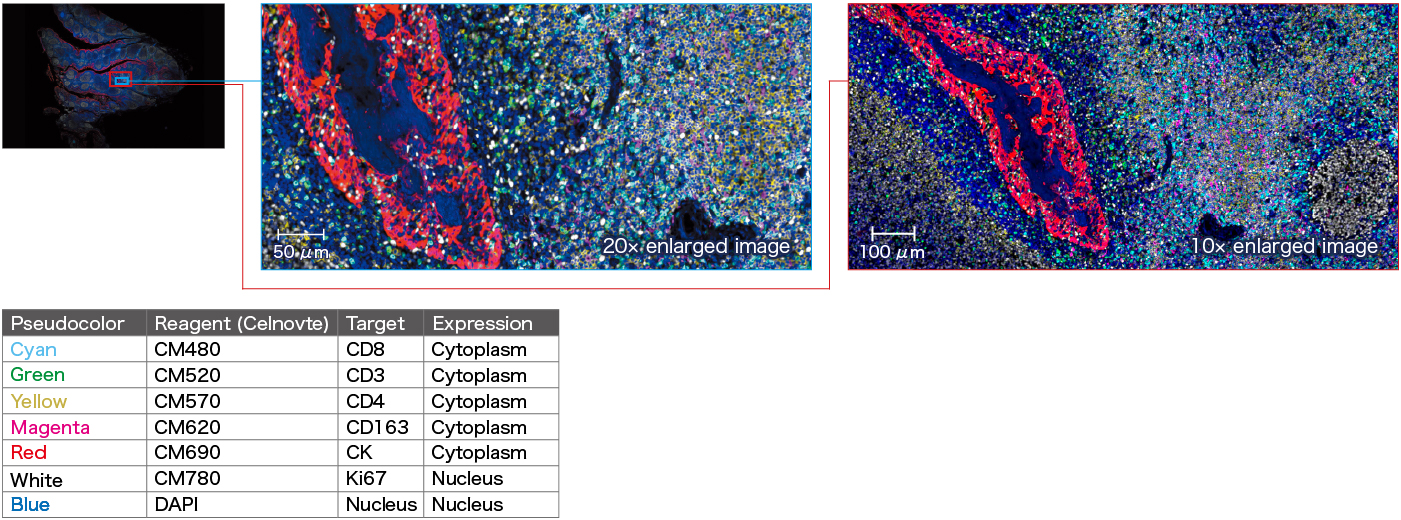
7-color multiplex fluorescence IHC detection kit
Six-plex overlay image of paraffin-embedded tonsil tissue sample
Vizgen “InSituPlex®” 4-plex OmniVUE™ Panels
4-plex overlay image of paraffin-embedded tonsil tissue sample
Staining principle of InSituPlex®
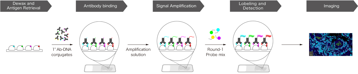
Cleavable Tyramide (TSA) Technology
7-plex overlay image of paraffin-embedded human tonsil sample
Cleavable Tyramide (TSA) Technology
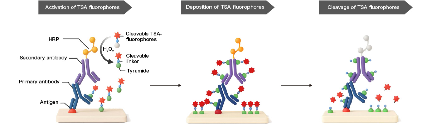
Cleavable Fluorescent Probe (CFP) - Spatomics’ Exclusive and Original Spatial Multiplexing Technology
- Proprietary cleavable TSA fluorophores designed for iterative staining & imaging
- Compatible with standard, off-the-shelf antibodies
- Best-in-class sensitivity and fluorescent dye selection
- Efficient fluorophore cleavage without loss of protein antigeneity
- Significantly reduce costs and development cycles for multiplex protein assays
- Compatible with on market RNA ISH assays for protein-RNA co-detection
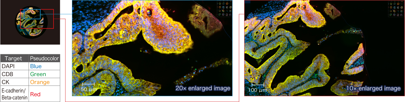
NaveniFlex™ Tissue Kit
4-plex overlay image of paraffin-embedded human colon sample
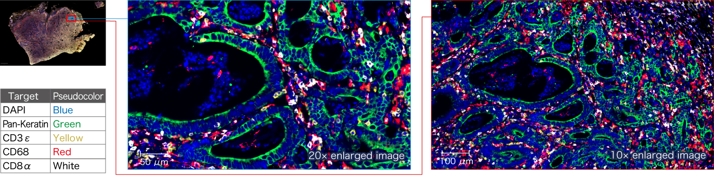
SignalStar® Multiplex IHC Kit
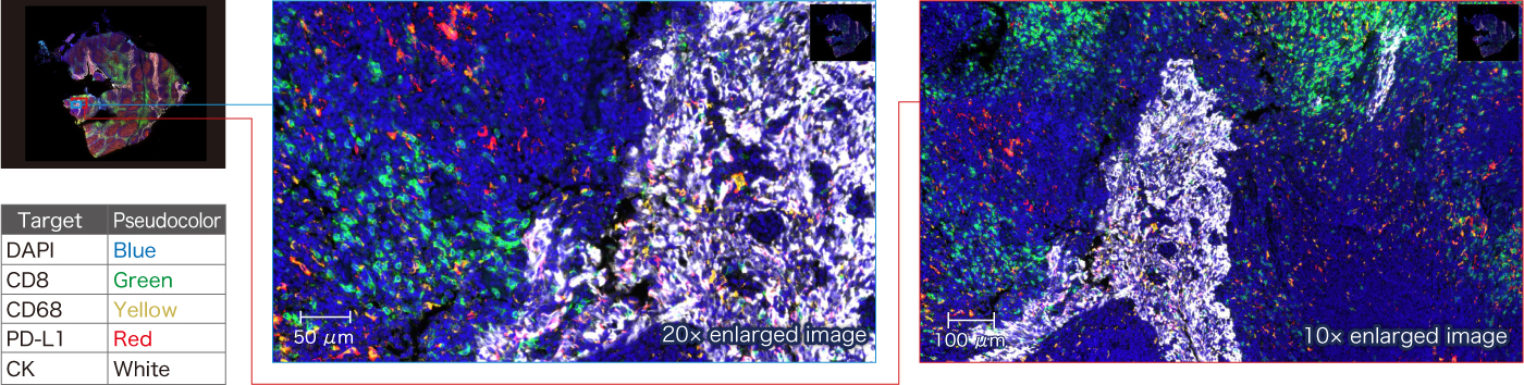
5-plex overlay image of paraffin-embedded human gastric cancer tissue
Overlay image of DAPI (blue) and individual targets (white)

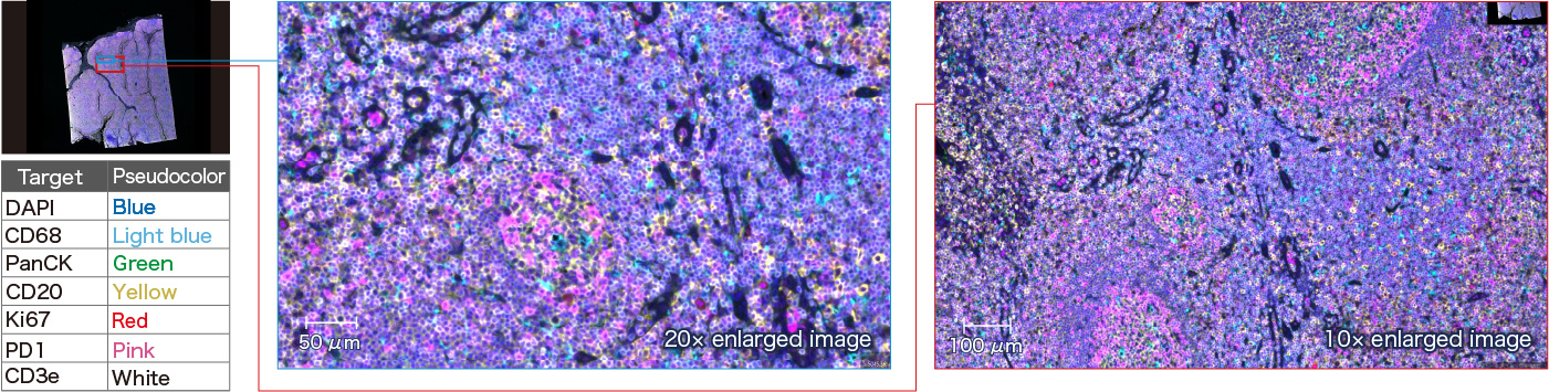
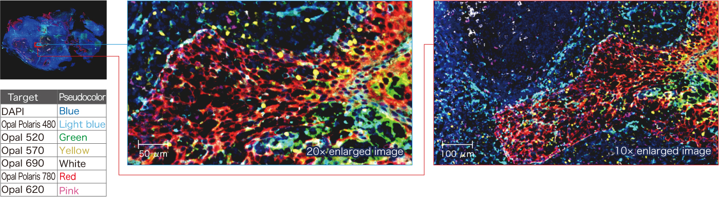
MOTiF PD-1/PD-L1 Panel 7-plex Kit
7-plex overlay image of paraffin-embedded human tonsil sample
Compatible with BF
The MoxiePlex allows side-by-side visualization of brightfield and fluorescence images within the MoxiePlex software. It captures high-resolution images of H&E-stained, immunohistochemically (IHC)-stained, and specially stained samples, providing a comprehensive view of tissue morphology for in-depth observation and analysis.
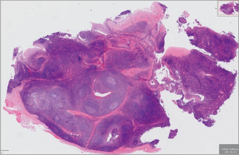
H&E
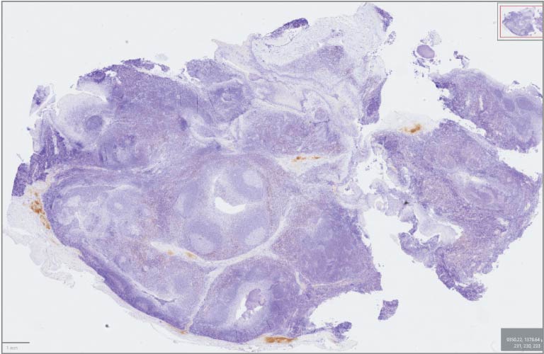
IHC
Automated sample recognition and exposure time calculation
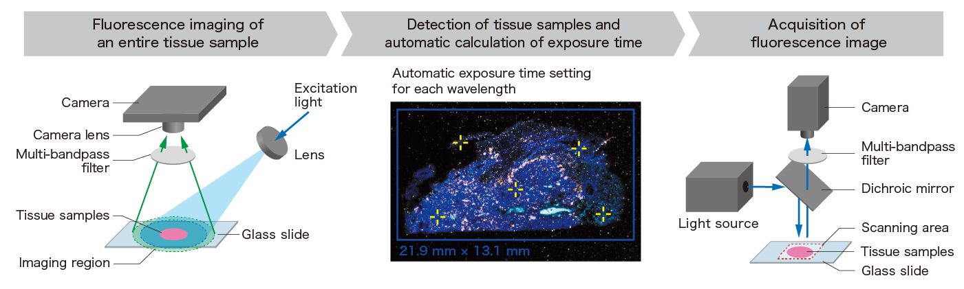
The MoxiePlex uses proprietary fluorescence optics technology and algorithms to automate sample recognition, scan range setting, focus position setting, and exposure time calculation. This not only reduces the manual workload required for setup, but also improves reproducibility when performing repeated measurements under the same conditions. Users can choose between fully automated operation to minimize manual effort or manual adjustments for greater flexibility and control. Furthermore, unmixing processing can be performed simultaneously with scanning, enabling efficient transition from data acquisition to analysis.
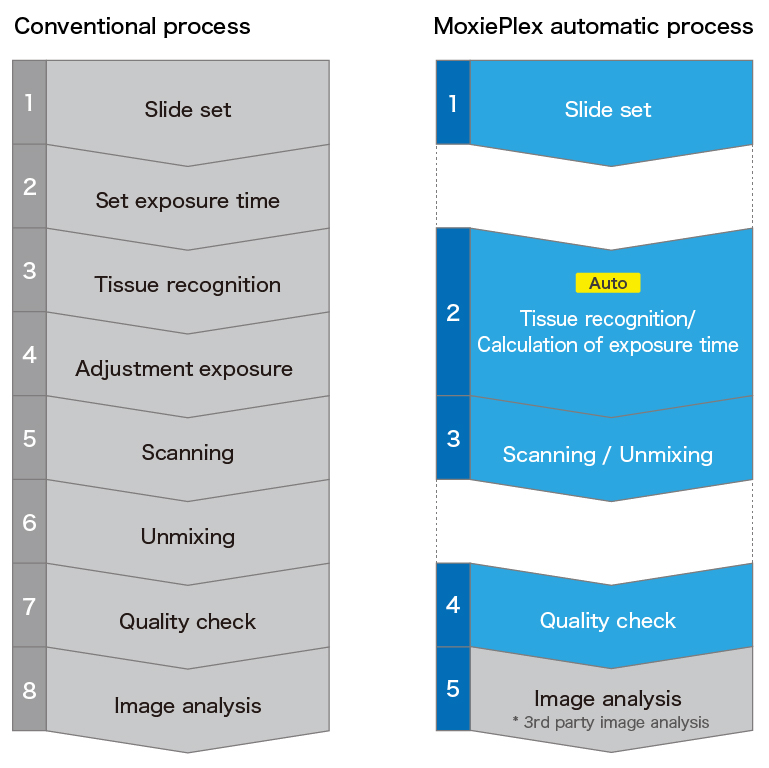
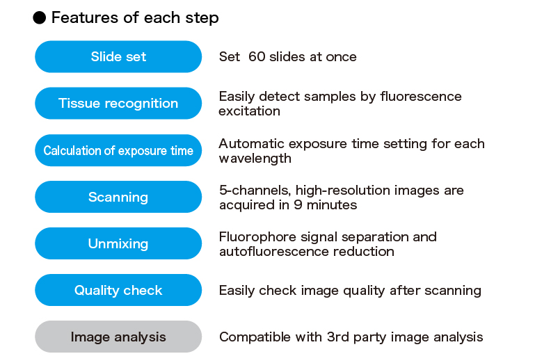
Proprietary fluorescence optical technology enabling automation
In MoxiePlex, the entire tissue sample is first imaged using low magnification and a wide field of view prior to scanning.
Based on these images, the system automatically detects the sample, adjusts exposure time, and determines optimal focus positions, enabling automation of processes that traditionally required manual adjustment. (Patent pending)
Unmixing technology for multiplex fluorescence analysis
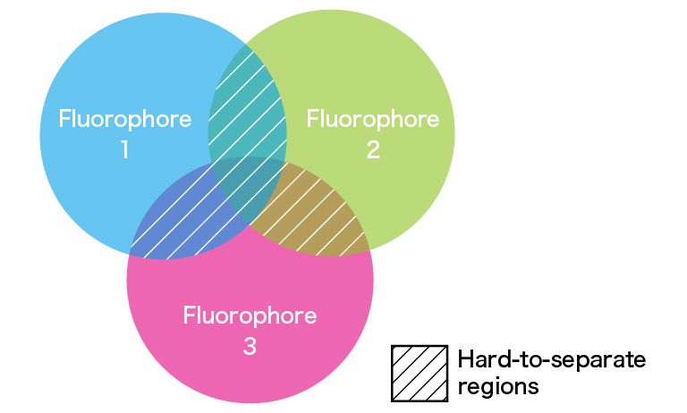
Unmixing in multiplex fluorescence staining is an analytical method that separates overlapping fluorescence signals into their individual components.
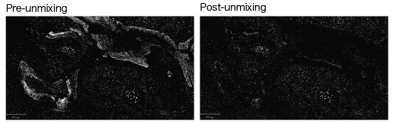
By applying unmixing techniques, MoxiePlex enables accurate extraction of target fluorophore signals while maintaining quantitative fluorescence intensity, even in multiplex-stained samples where spectral overlap and crosstalk are prevalent.
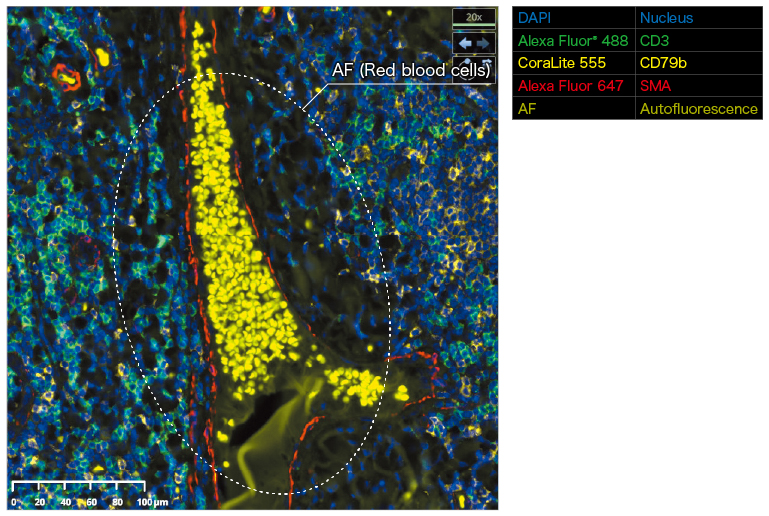
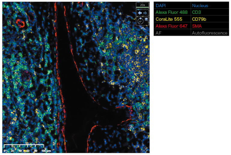
Additionally, this approach reduces autofluorescence components with broad emission spectra, such as those originating from red blood cells, contributing to improved signal separation performance and enhanced analytical accuracy in multiplex fluorescence analysis.
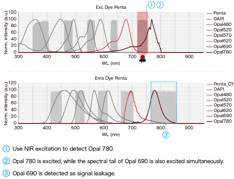
When detecting Opal 780
Library creation for enhanced unmixing accuracy
MoxiePlex is equipped with a library creation function that enables the generation of libraries used for unmixing from pre-acquired images of single-stained samples.
In addition, for samples exhibiting autofluorescence, libraries including autofluorescence components can also be created, enabling more accurate unmixing analysis.
The system also allows flexible updating of libraries in response to changes in assay conditions or imaging systems, helping to reduce operational costs and processing time associated with unmixing analysis.
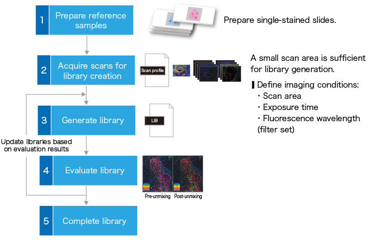
MoxiePlex library creation workflow
Application examples
Evaluation of a multiplex fluorescence–stained sample (7-plex)
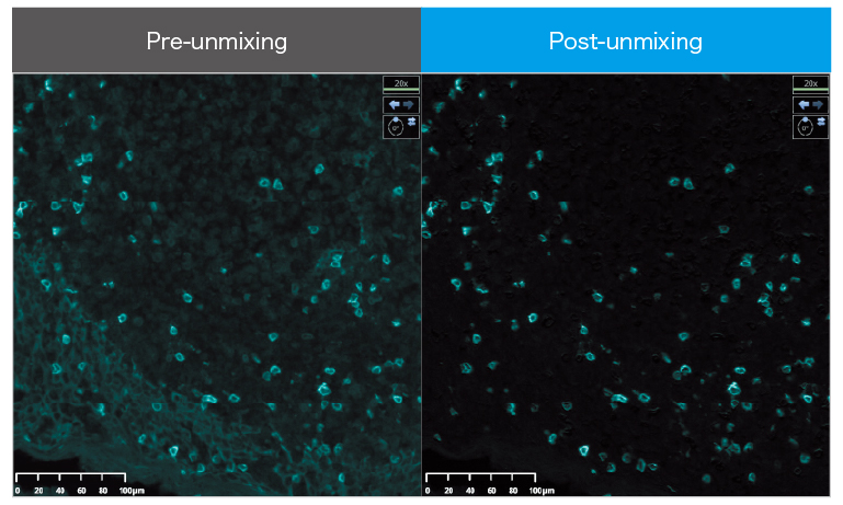
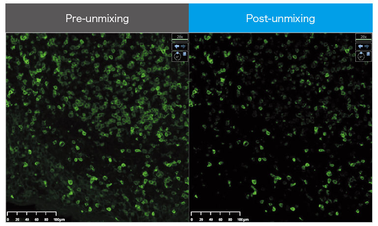
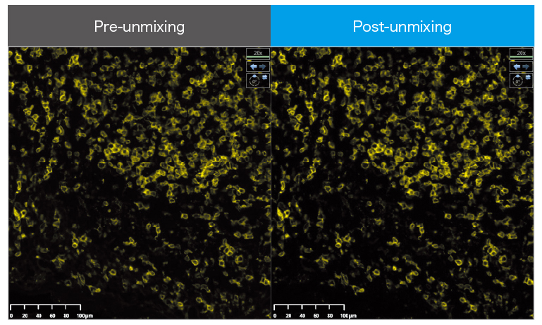
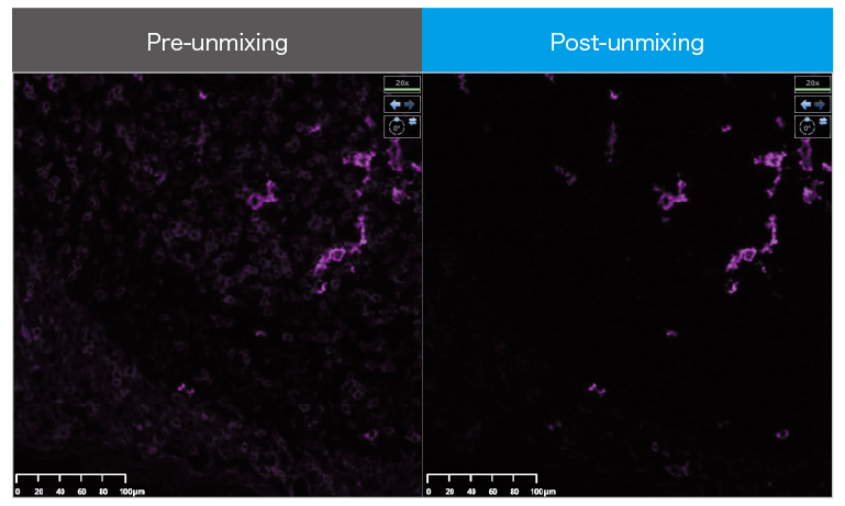
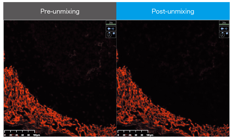
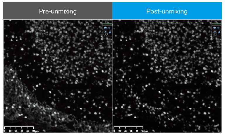
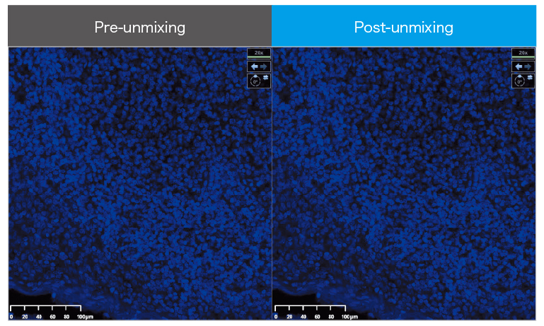
Autofluorescence
Related documents
Technical note
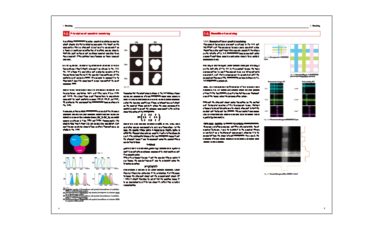
In multiplex fluorescence imaging, signal crosstalk between fluorophores and tissue autofluorescence can compromise analytical accuracy. This technical note explains the principles and benefits of the unmixing technology incorporated in the MoxiePlex Multispectral Imaging System, with practical examples demonstrating its effectiveness. It also introduces key features that support high-precision analysis and streamlined workflows, including compatibility with a wide range of spatial biology assays and fluorescence macro functionality for automated imaging optimization.
An interview article published on the website of the scientific journal Nature.
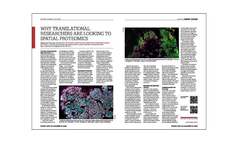
This article is an interview published on the website of the world‑renowned scientific journal Nature. In this discussion, three experts—Dr. Carlo Bifulco (Providence Genomics), Dr. David Rimm (Yale University), and Don Ariyakumar (HAMAMATSU CORPORATION)—share their perspectives on the clinical application of spatial proteomics technologies, which enable detailed analysis of proteins within cancer cells and their surrounding microenvironment. They also discuss the unique features of MoxiePlex and its contributions to diagnostics and therapeutic decision‑making.
Related videos
SITC 2025 - From Spatial Biology to Clinical Utility: Advancing Lung Cancer Diagnostics
The SITC 2025 session, “From Spatial Biology to Clinical Utility: Advancing Lung Cancer Diagnostics,” explores cutting-edge advancements in spatial biology technologies and their transformative potential in improving lung cancer diagnosis and treatment strategies. The discussion features insights from Dr. Carlo Bifulco, Chief Medical Officer at Providence Genomics and Director of Translational Molecular Pathology at EACRI, and Xiaoshan Wang, VP of Business Development for Spatomics, and who share perspectives from both industry and clinical research.
From 1:40|Maximizing Diagnostic Power from Limited Lung Cancer Tissue
Carlo Bifulco, MD – Chief Medical Officer at Providence Genomics | Director of Translational Molecular Pathology at the Earle A Chiles Research Institute
From 34:43|Breaking the Multiplex Barrier with CFP™ Cleavable Tyramide Technology
Xiaoshan Wang – Spatomics
Specifications
| Product name | MoxiePlex Multi-spectral imaging system | |
|---|---|---|
| Product number | C16919-01 | |
| Cassette loader | Up to 60 slides | |
| Compatible glass slide | 75.0 mm to 76.0 mm × 25.0 mm to 26.0 mm (Thickness: 0.9 mm to 1.2 mm) |
|
| Objective lens | 20× NA 0.8 |
|
| Scanning resolution | 20x mode | 0.46 μm/pixel |
| 40x mode | 0.23 μm/pixel | |
| Brightfield scan speed | 20x mode | Approx. 60 s(15 mm × 15 mm) |
| 40x mode | Approx. 70 s(15 mm × 15 mm) | |
| Fluorescence throughput *1 | 20x mode | Approx. 12 min.(15 mm × 15 mm, 5 channels) |
| 40x mode | Approx. 14 min.(15 mm × 15 mm, 5 channels) | |
| Fluorescence camera | In-house CMOS camera | |
| Z-stack feature | Included | |
| Image format | OME-TIFF (16 bit, 8 bit), NDPI (8 bit) | |
| Power supply | AC 100 V to AC 240 V | |
| Power consumption | Approx. 180 VA | |
*1 Fluorescence throughput: The time from loading the glass slide, macro photography, prefocusing 5 points, scanning, image processing, to unloading the glass slide.
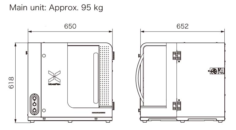
Dimensions
- Confirmation
-
It looks like you're in the . If this is not your location, please select the correct region or country below.
You're headed to Hamamatsu Photonics website for US (English). If you want to view an other country's site, the optimized information will be provided by selecting options below.
In order to use this website comfortably, we use cookies. For cookie details please see our cookie policy.
- Cookie Policy
-
This website or its third-party tools use cookies, which are necessary to its functioning and required to achieve the purposes illustrated in this cookie policy. By closing the cookie warning banner, scrolling the page, clicking a link or continuing to browse otherwise, you agree to the use of cookies.
Hamamatsu uses cookies in order to enhance your experience on our website and ensure that our website functions.
You can visit this page at any time to learn more about cookies, get the most up to date information on how we use cookies and manage your cookie settings. We will not use cookies for any purpose other than the ones stated, but please note that we reserve the right to update our cookies.
1. What are cookies?
For modern websites to work according to visitor’s expectations, they need to collect certain basic information about visitors. To do this, a site will create small text files which are placed on visitor’s devices (computer or mobile) - these files are known as cookies when you access a website. Cookies are used in order to make websites function and work efficiently. Cookies are uniquely assigned to each visitor and can only be read by a web server in the domain that issued the cookie to the visitor. Cookies cannot be used to run programs or deliver viruses to a visitor’s device.
Cookies do various jobs which make the visitor’s experience of the internet much smoother and more interactive. For instance, cookies are used to remember the visitor’s preferences on sites they visit often, to remember language preference and to help navigate between pages more efficiently. Much, though not all, of the data collected is anonymous, though some of it is designed to detect browsing patterns and approximate geographical location to improve the visitor experience.
Certain type of cookies may require the data subject’s consent before storing them on the computer.
2. What are the different types of cookies?
This website uses two types of cookies:
- First party cookies. For our website, the first party cookies are controlled and maintained by Hamamatsu. No other parties have access to these cookies.
- Third party cookies. These cookies are implemented by organizations outside Hamamatsu. We do not have access to the data in these cookies, but we use these cookies to improve the overall website experience.
3. How do we use cookies?
This website uses cookies for following purposes:
- Certain cookies are necessary for our website to function. These are strictly necessary cookies and are required to enable website access, support navigation or provide relevant content. These cookies direct you to the correct region or country, and support security and ecommerce. Strictly necessary cookies also enforce your privacy preferences. Without these strictly necessary cookies, much of our website will not function.
- Analytics cookies are used to track website usage. This data enables us to improve our website usability, performance and website administration. In our analytics cookies, we do not store any personal identifying information.
- Functionality cookies. These are used to recognize you when you return to our website. This enables us to personalize our content for you, greet you by name and remember your preferences (for example, your choice of language or region).
- These cookies record your visit to our website, the pages you have visited and the links you have followed. We will use this information to make our website and the advertising displayed on it more relevant to your interests. We may also share this information with third parties for this purpose.
Cookies help us help you. Through the use of cookies, we learn what is important to our visitors and we develop and enhance website content and functionality to support your experience. Much of our website can be accessed if cookies are disabled, however certain website functions may not work. And, we believe your current and future visits will be enhanced if cookies are enabled.
4. Which cookies do we use?
There are two ways to manage cookie preferences.
- You can set your cookie preferences on your device or in your browser.
- You can set your cookie preferences at the website level.
If you don’t want to receive cookies, you can modify your browser so that it notifies you when cookies are sent to it or you can refuse cookies altogether. You can also delete cookies that have already been set.
If you wish to restrict or block web browser cookies which are set on your device then you can do this through your browser settings; the Help function within your browser should tell you how. Alternatively, you may wish to visit www.aboutcookies.org, which contains comprehensive information on how to do this on a wide variety of desktop browsers.
5. What are Internet tags and how do we use them with cookies?
Occasionally, we may use internet tags (also known as action tags, single-pixel GIFs, clear GIFs, invisible GIFs and 1-by-1 GIFs) at this site and may deploy these tags/cookies through a third-party advertising partner or a web analytical service partner which may be located and store the respective information (including your IP-address) in a foreign country. These tags/cookies are placed on both online advertisements that bring users to this site and on different pages of this site. We use this technology to measure the visitors' responses to our sites and the effectiveness of our advertising campaigns (including how many times a page is opened and which information is consulted) as well as to evaluate your use of this website. The third-party partner or the web analytical service partner may be able to collect data about visitors to our and other sites because of these internet tags/cookies, may compose reports regarding the website’s activity for us and may provide further services which are related to the use of the website and the internet. They may provide such information to other parties if there is a legal requirement that they do so, or if they hire the other parties to process information on their behalf.
If you would like more information about web tags and cookies associated with on-line advertising or to opt-out of third-party collection of this information, please visit the Network Advertising Initiative website http://www.networkadvertising.org.
6. Analytics and Advertisement Cookies
We use third-party cookies (such as Google Analytics) to track visitors on our website, to get reports about how visitors use the website and to inform, optimize and serve ads based on someone's past visits to our website.
You may opt-out of Google Analytics cookies by the websites provided by Google:
https://tools.google.com/dlpage/gaoptout?hl=en
As provided in this Privacy Policy (Article 5), you can learn more about opt-out cookies by the website provided by Network Advertising Initiative:
http://www.networkadvertising.org
We inform you that in such case you will not be able to wholly use all functions of our website.
Close