![]()
Products


We are actively taking measures to improve product quality levels.
Applications


Why Hamamatsu?
Support


Our company


Investors


Japan (EN)
Select your region or country.

Fluorescence imaging
What is fluorescence imaging?
Fluorescence imaging is an imaging technique that visualizes cells and molecules by fluorescently labeling them using fluorescent dyes, fluorescent proteins, and NV centers. Generally, cells and molecules are colorless and transparent, so they cannot be observed as they are. There are various staining methods other than fluorescent labeling to visualize cells; however, many of them can damage the cells. Fluorescent dyes, fluorescent proteins, and NV centers used in fluorescence imaging can label cells while they are alive, enabling live cell imaging of organisms. This allows us to obtain various information such as cell structure and function, and molecular interactions. Fluorescence imaging has become an important tool in the development of biology.
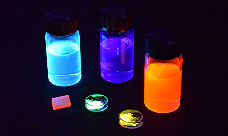
Fluorescence imaging made significant advancements thanks to the Nobel Prize in Chemistry awarded in 2008 to three researchers: Professor Osamu Shimomura, Professor Martin Chalfie, and Professor Roger Y. Tsien. The prize recognized their discovery and development of the green fluorescent protein (GFP). GFP is a protein isolated from the bioluminescent jellyfish. Professor Shimomura was the first to successfully isolate GFP from jellyfish after observing that it emits green light when exposed to ultraviolet radiation. Subsequently, Professor Martin Chalfie contributed to understanding the fluorescence emission mechanism and successfully expressed GFP within living cells using genetic engineering techniques. Additionally, Professor Roger Y. Tsien elucidated the molecular mechanism of GFP chromophore formation, allowing the creation of artificial fluorescent proteins that emit various colors beyond green. This breakthrough enabled the expression of different colored fluorescent proteins within living organisms, facilitating simultaneous tracking of multiple biological phenomena, including the analysis of interactions between different proteins.
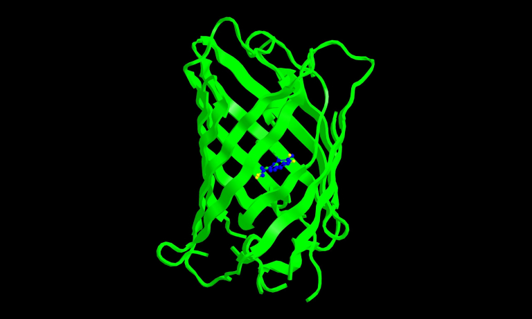
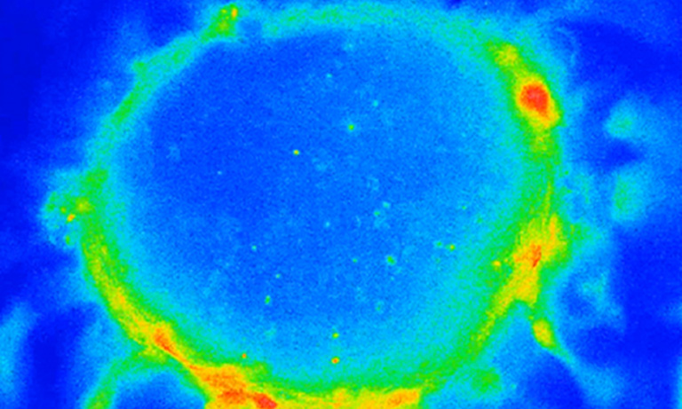
Figure 1: Structure of green fluorescent protein (GFP)
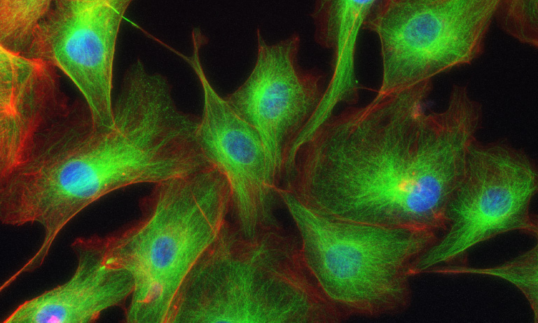
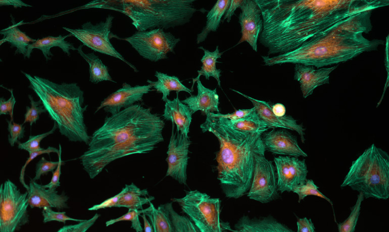
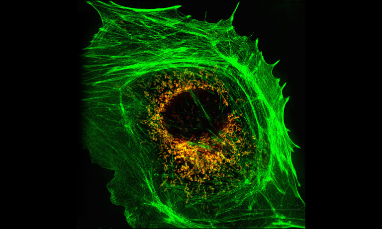
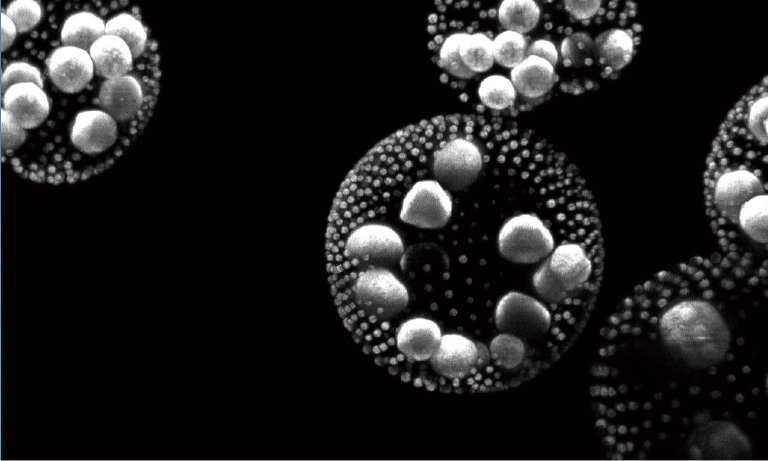
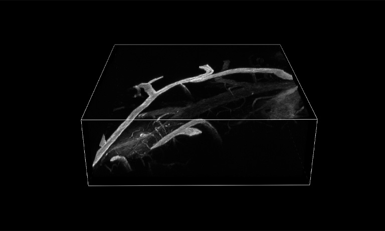
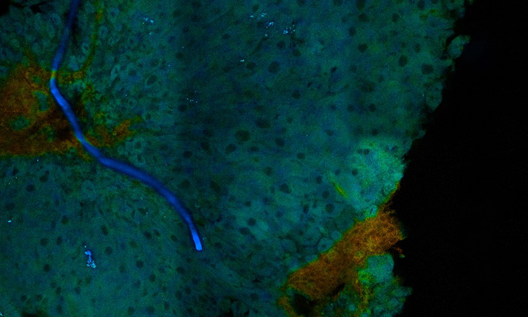
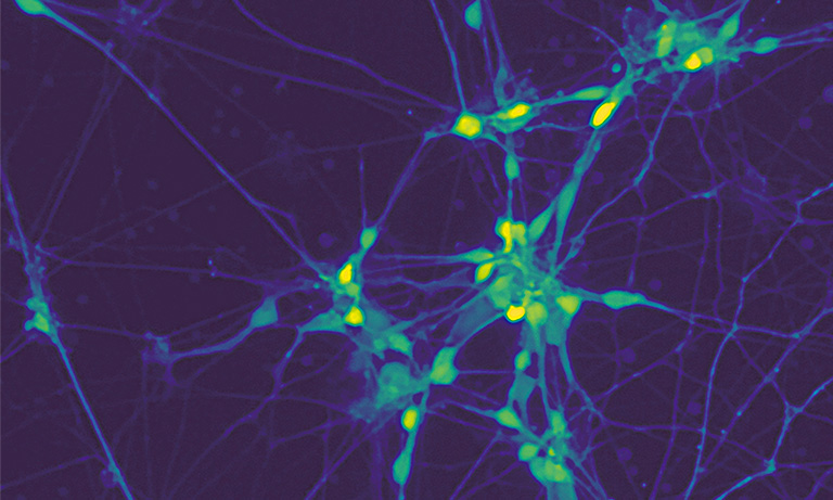
Figure 2: Example of fluorescence-stained cells
What is fluorescence microscopy?
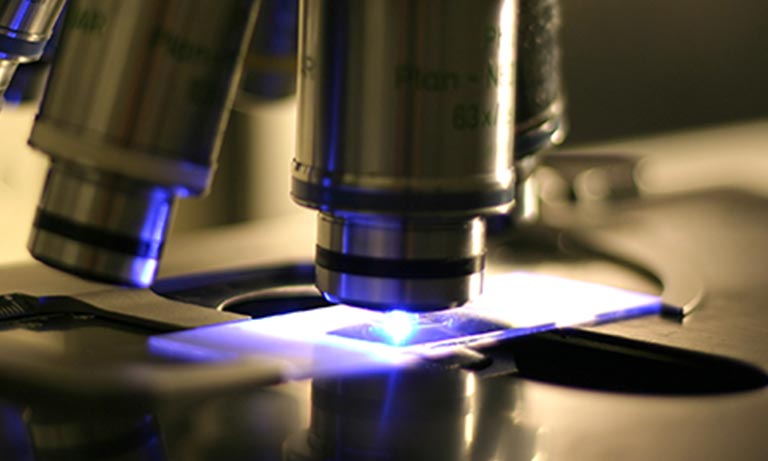
A fluorescence microscope is a type of microscope used for imaging cellular structures through fluorescence. A sample labeled with a fluorescent dye or protein is irradiated with excitation light, and the fluorescence emitted from the sample is focused by an objective lens and photographed by a camera.
A fluorescence microscope mainly consists of an optical system including a light source, filter, dichroic mirror, and objective lens. Figure 3 shows a schematic diagram of a commonly used epifluorescence microscope. In an epifluorescence microscope fluorescence microscope, light emitted from the light source (excitation light) has its unwanted wavelengths cut off by an excitation filter. The excitation light is then reflected by a dichroic mirror and irradiated onto the sample through an objective lens. The sample irradiated by the excitation light emits fluorescence, which is passed through the objective lens, dichroic mirror, and fluorescence filter (absorption filter) before being guided to a detector such as an eyepiece lens or camera.
In recent years, various fluorescence imaging techniques have been developed, including confocal microscopy, super-resolution microscopy, light-sheet microscopy, and multiphoton microscopy. These fluorescence imaging techniques enable observation at higher resolution, faster speeds, and greater depths than ever before.
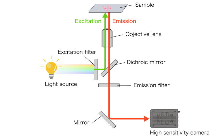
Figure 3: Schematic diagram of an epifluorescence microscope
Find examples and products from microscopy techniques
An imaging technique that removes fluorescence generated outside the focal plane, resulting in high-resolution images with minimal blur.
An imaging technique that is used to observe with resolution beyond the diffraction limit of conventional optical microscopes.
An imaging technique that allows rapid image acquisition while minimizing phototoxicity by exciting the sample using sheet illumination.
An imaging technique that enables deep image acquisition by utilizing multiphoton excitation phenomena.
An imaging technique that maps the spatial distribution of sample fluorescence lifetimes and acquires images.
Find examples and products from experimental methods
Related products
Detectors
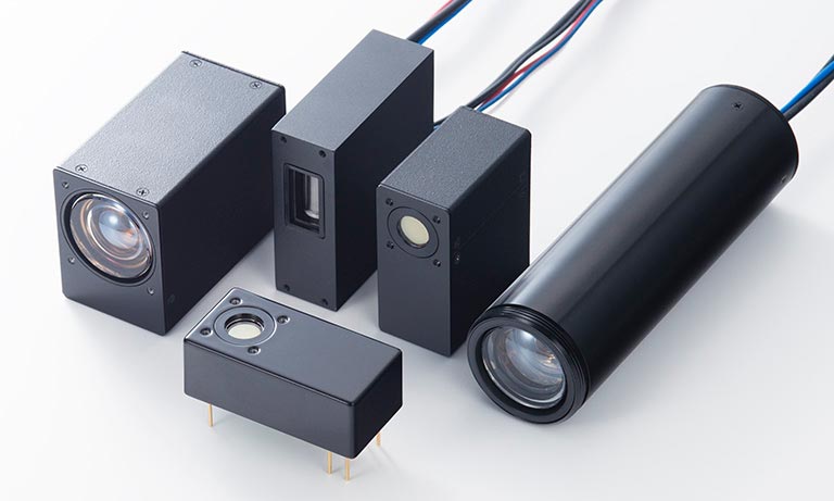
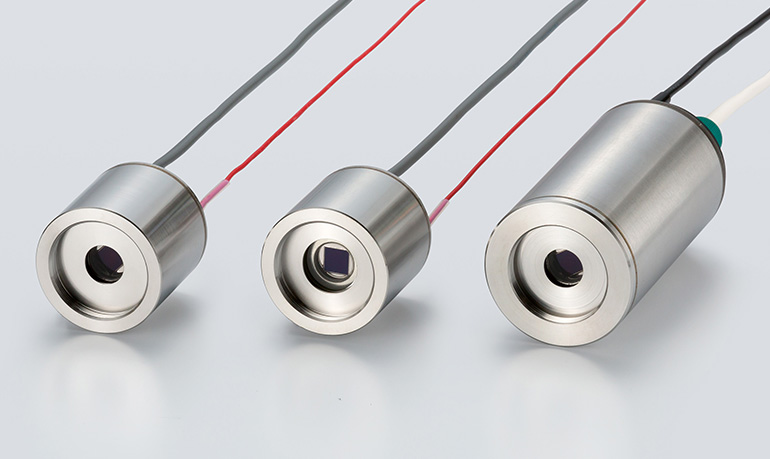
PMT modules are comprised of a photomultiplier tube to convert light into electrical signals, a high-voltage power supply circuit, and a voltage divider circuit to distribute the optimum voltage to each dynode.
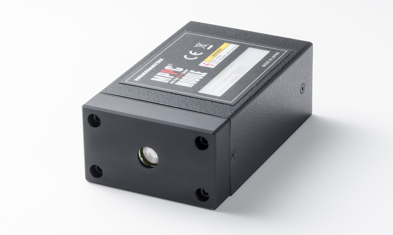
Modules (SiPM modules) that contain an MPPC capable of detecting extremely low-level light.
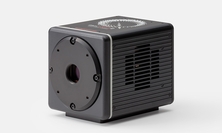
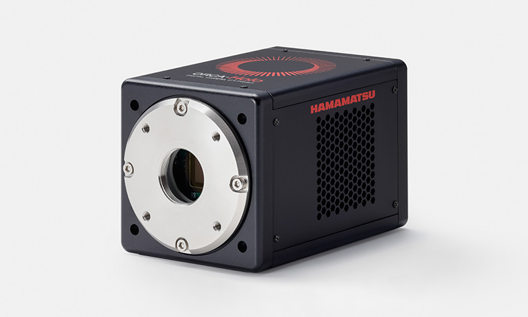
The world's first camera to incorporate the qCMOS image sensor.
The camera achieves the ultimate in quantitative imaging.
HPDs are completely new PMTs that contain a semiconductor device within a vacuum or electron tube, enabling efficient electron multiplication with less noise.
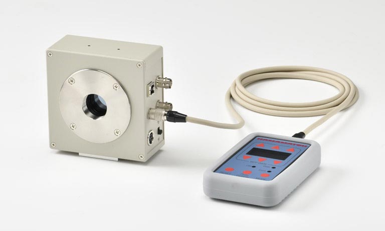
High-speed gated II units use gate operation to capture instantaneous images of high-speed phenomena occurring within extremely short time durations.
Light source
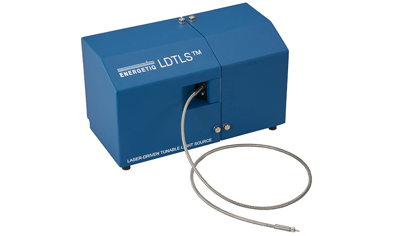
The Laser-Driven Tunable Light Source (LDTLS™) is a compact, fully integrated and highly stable tunable broadband light source based on proven Laser-Driven Light Source (LDLS™) technology.
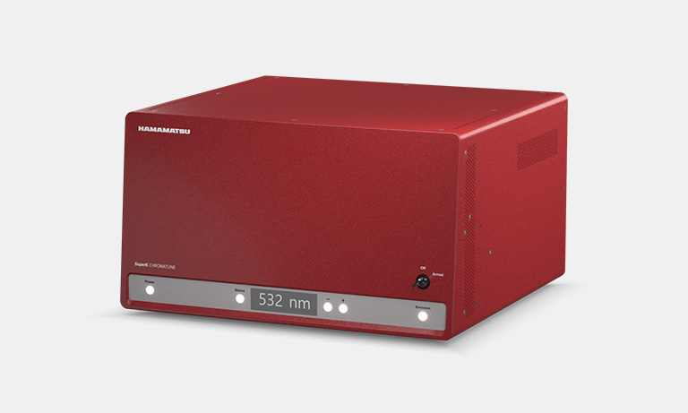
The SuperK CHROMATUNE is the World’s broadest tunable laser giving you an unmatched 400-1000 nm tuning range.
Others
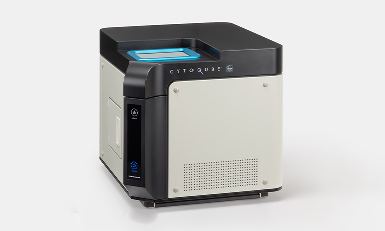
CYTOQUBE is a Light-Sheet Microplate Cytometer that performs high-speed fluorescence imaging and analysis for both 2D and 3D cell-cultured samples.
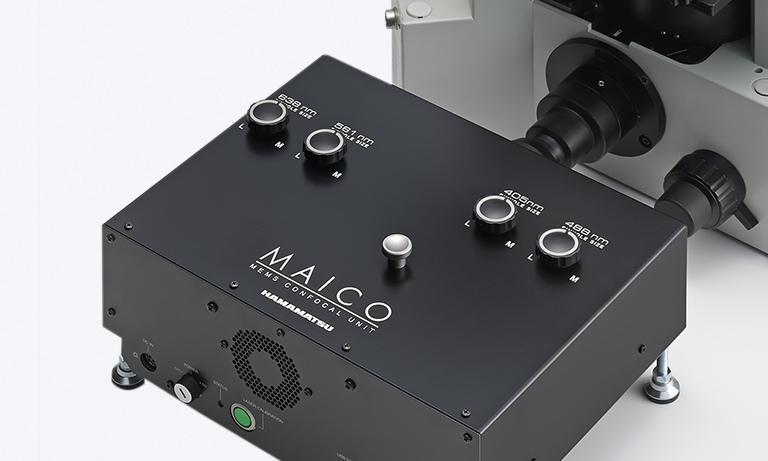
Simply attach this MEMS confocal unit to an inverted microscope and enable confocal fluorescence imaging.
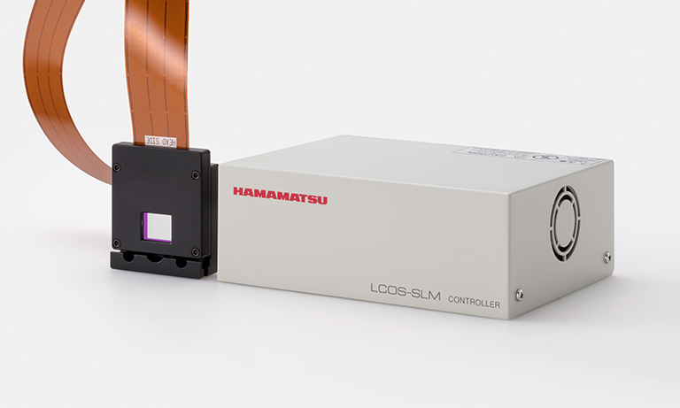
LCOS-SLM are reflective spatial light phase modulators that freely modulates optical phases and optical phase of laser is modulated by the liquid crystal.
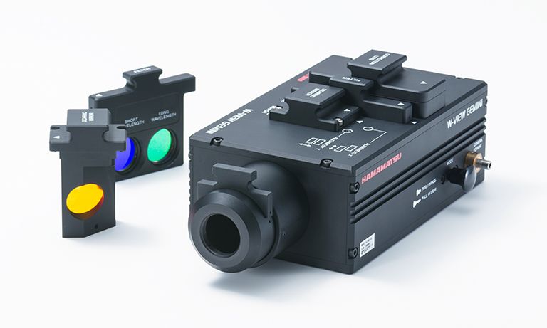
Image splitting optics provide one pair of dual wavelength images to cameras.
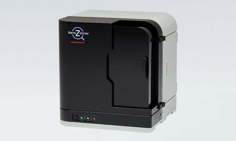
Digital slide scanner is a family of whole slide scanners that convert glass slides into high-resolution digital data by high-speed scanning.
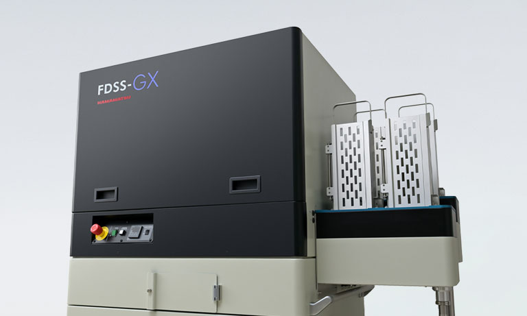
High speed, kinetic plate imager for fluorescence / luminescence assays in GPCR / ion channel drug discovery field.
- Confirmation
-
It looks like you're in the . If this is not your location, please select the correct region or country below.
You're headed to Hamamatsu Photonics website for JP (English). If you want to view an other country's site, the optimized information will be provided by selecting options below.
In order to use this website comfortably, we use cookies. For cookie details please see our cookie policy.
- Cookie Policy
-
This website or its third-party tools use cookies, which are necessary to its functioning and required to achieve the purposes illustrated in this cookie policy. By closing the cookie warning banner, scrolling the page, clicking a link or continuing to browse otherwise, you agree to the use of cookies.
Hamamatsu uses cookies in order to enhance your experience on our website and ensure that our website functions.
You can visit this page at any time to learn more about cookies, get the most up to date information on how we use cookies and manage your cookie settings. We will not use cookies for any purpose other than the ones stated, but please note that we reserve the right to update our cookies.
1. What are cookies?
For modern websites to work according to visitor’s expectations, they need to collect certain basic information about visitors. To do this, a site will create small text files which are placed on visitor’s devices (computer or mobile) - these files are known as cookies when you access a website. Cookies are used in order to make websites function and work efficiently. Cookies are uniquely assigned to each visitor and can only be read by a web server in the domain that issued the cookie to the visitor. Cookies cannot be used to run programs or deliver viruses to a visitor’s device.
Cookies do various jobs which make the visitor’s experience of the internet much smoother and more interactive. For instance, cookies are used to remember the visitor’s preferences on sites they visit often, to remember language preference and to help navigate between pages more efficiently. Much, though not all, of the data collected is anonymous, though some of it is designed to detect browsing patterns and approximate geographical location to improve the visitor experience.
Certain type of cookies may require the data subject’s consent before storing them on the computer.
2. What are the different types of cookies?
This website uses two types of cookies:
- First party cookies. For our website, the first party cookies are controlled and maintained by Hamamatsu. No other parties have access to these cookies.
- Third party cookies. These cookies are implemented by organizations outside Hamamatsu. We do not have access to the data in these cookies, but we use these cookies to improve the overall website experience.
3. How do we use cookies?
This website uses cookies for following purposes:
- Certain cookies are necessary for our website to function. These are strictly necessary cookies and are required to enable website access, support navigation or provide relevant content. These cookies direct you to the correct region or country, and support security and ecommerce. Strictly necessary cookies also enforce your privacy preferences. Without these strictly necessary cookies, much of our website will not function.
- Analytics cookies are used to track website usage. This data enables us to improve our website usability, performance and website administration. In our analytics cookies, we do not store any personal identifying information.
- Functionality cookies. These are used to recognize you when you return to our website. This enables us to personalize our content for you, greet you by name and remember your preferences (for example, your choice of language or region).
- These cookies record your visit to our website, the pages you have visited and the links you have followed. We will use this information to make our website and the advertising displayed on it more relevant to your interests. We may also share this information with third parties for this purpose.
Cookies help us help you. Through the use of cookies, we learn what is important to our visitors and we develop and enhance website content and functionality to support your experience. Much of our website can be accessed if cookies are disabled, however certain website functions may not work. And, we believe your current and future visits will be enhanced if cookies are enabled.
4. Which cookies do we use?
There are two ways to manage cookie preferences.
- You can set your cookie preferences on your device or in your browser.
- You can set your cookie preferences at the website level.
If you don’t want to receive cookies, you can modify your browser so that it notifies you when cookies are sent to it or you can refuse cookies altogether. You can also delete cookies that have already been set.
If you wish to restrict or block web browser cookies which are set on your device then you can do this through your browser settings; the Help function within your browser should tell you how. Alternatively, you may wish to visit www.aboutcookies.org, which contains comprehensive information on how to do this on a wide variety of desktop browsers.
5. What are Internet tags and how do we use them with cookies?
Occasionally, we may use internet tags (also known as action tags, single-pixel GIFs, clear GIFs, invisible GIFs and 1-by-1 GIFs) at this site and may deploy these tags/cookies through a third-party advertising partner or a web analytical service partner which may be located and store the respective information (including your IP-address) in a foreign country. These tags/cookies are placed on both online advertisements that bring users to this site and on different pages of this site. We use this technology to measure the visitors' responses to our sites and the effectiveness of our advertising campaigns (including how many times a page is opened and which information is consulted) as well as to evaluate your use of this website. The third-party partner or the web analytical service partner may be able to collect data about visitors to our and other sites because of these internet tags/cookies, may compose reports regarding the website’s activity for us and may provide further services which are related to the use of the website and the internet. They may provide such information to other parties if there is a legal requirement that they do so, or if they hire the other parties to process information on their behalf.
If you would like more information about web tags and cookies associated with on-line advertising or to opt-out of third-party collection of this information, please visit the Network Advertising Initiative website http://www.networkadvertising.org.
6. Analytics and Advertisement Cookies
We use third-party cookies (such as Google Analytics) to track visitors on our website, to get reports about how visitors use the website and to inform, optimize and serve ads based on someone's past visits to our website.
You may opt-out of Google Analytics cookies by the websites provided by Google:
https://tools.google.com/dlpage/gaoptout?hl=en
As provided in this Privacy Policy (Article 5), you can learn more about opt-out cookies by the website provided by Network Advertising Initiative:
http://www.networkadvertising.org
We inform you that in such case you will not be able to wholly use all functions of our website.
Close