![]()
Products


We are actively taking measures to improve product quality levels.
Applications


Why Hamamatsu?
Resources


Support


Our company


Investors


United States (JA)
国・地域を選択してください。
Features / Clinical Usefulness / Safety
What is a PET scan?
What kind of examination is a PET scan?
PET is an abbreviation for Positron Emission Tomography. In Japanese, it is referred to as “Yōdenshi Dansō Satsueihō” (positron emission tomography). A PET scan is an imaging technique used to visualize the function of organs such as the heart and brain in cross-sectional images, helping to identify the cause of disease and assess disease conditions. In a PET examination, a radiopharmaceutical suitable for the target of the examination is administered into the body. The PET scanner then images its distribution in the body, allowing visualization of cellular metabolism and function.
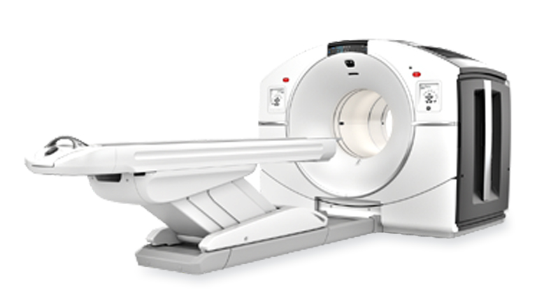
PET scanner Discovery MI
Why can PET scans detect cancer?
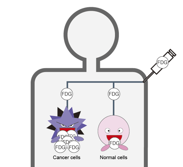
Cancer cells divide more actively than normal cells and therefore require a much larger amount of glucose as an energy source. For this reason, a radiopharmaceutical called FDG—a substance with properties similar to glucose and labeled with a small amount of a radioactive isotope—is administered intravenously. FDG accumulates more abundantly in cancer cells than in normal cells.Using a PET scanner, the distribution of FDG in the body is imaged, allowing the location and size of cancer to be visualized.
Right image: Because cancer cells require more glucose than normal cells, a large amount of FDG accumulates in cancer cells.
How does cancer appear on a PET scan?
The degree of FDG accumulation is displayed as differences in brightness on black-and-white images and as differences in color on color images. Physiological FDG uptake is normally seen in actively functioning tissues such as brain neurons and heart muscle. Because FDG is excreted in the urine, accumulation is also observed in the kidneys, ureters, and bladder. In addition, FDG may accumulate in inflammatory cells and benign tumors. Therefore, not all areas showing FDG uptake indicate cancer.
Features of PET scans
A wide area can be examined in a single session
A PET scan typically images a wide area of the body, from the head to the pelvis, in a single examination. This makes it useful for identifying the primary tumor and determining whether metastasis is present. In some cases, cancer may be detected in unexpected locations that are not usually examined in routine health screenings.
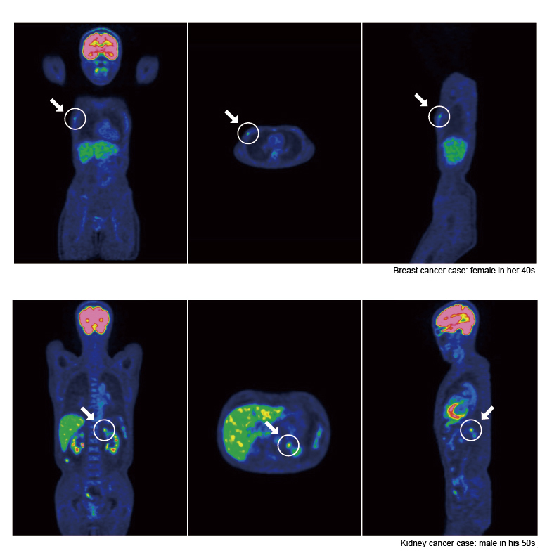
Image provided by Hamamatsu PET Diagnostic Center
Useful for early detection of cancer
PET scans provide information about cellular activity and metabolism. When combined with structural information obtained from conventional imaging methods
such as X‑ray CT or MRI, PET scans can reveal more detailed conditions of the body, making them effective for the diagnosis of early-stage cancer.
A minimally invasive examination
PET examination does not cause pain other than the injection. The scan is performed while wearing clothes, and it does not involve significant discomfort. There is no significant pain or discomfort.
Usefulness of PET examinations
PET examinations do not detect all cancers.
PET examinations are useful for cancer diagnosis and early detection; however, they are not effective for detecting all types of cancer. FDG normally accumulates in the kidneys and urinary bladder even in healthy individuals. For this reason, cancers in these organs, as well as prostate cancer located near the bladder, may be difficult to detect. In addition, conventional screening methods are more effective for certain cancers, such as early-stage gastric cancer and cervical cancer. FDG also accumulates in areas of inflammation and in benign tumors. FDG also accumulates in areas of inflammation and in benign tumors.
When PET is used for health screening purposes, it is important to undergo additional examinations—such as MRI, CT, and endoscopy—in combination, rather than relying solely on PET. As with other screening tests, PET examinations may yield false-positive results (interpreted as suspicious for cancer when no cancer is present) and false-negative results (interpreted as negative even when cancer is present). Furthermore, FDG uptake is affected by blood glucose levels; therefore, in patients with diabetes, images may be more difficult to interpret.
■ Usefulness of FDG-PET in cancer detection
| Cancers for which FDG-PET is considered highly useful | Cancers for which FDG-PET Is considered less useful |
|---|---|
|
* In PET-based cancer screening, it is recommended that PET be used in combination with other diagnostic examinations. |
Source: FDG-PET Cancer Screening Guidelines (3rd Edition),The Japanese Society of Nuclear Medicine / PET and Nuclear Medicine Subcommittee
Which cancer screening tests are considered effective?
Several cancer screening tests listed in the table below have been shown, through scientific studies, to reduce cancer mortality and are therefore considered effective as cancer screening methods. At the time of this report, lung cancer screening using X‑ray CT and prostate cancer screening using PSA testing are categorized as “under evaluation.” The effectiveness of PET cancer screening as a population-based screening test has not yet been confirmed.
■ The kind of cancer screening recommended by the government
| Type | Screening Items / Examination Items | Target age | Screening interval |
|---|---|---|---|
| gastric cancer | medical questionnaire + either gastric X‑ray examination or upper gastrointestinal endoscopy | 50 years and older | once every 2 years |
| cervical cancer | medical questionnaire + visual inspection + cervical cytology and pelvic examination | 20 years and older | once every 2 years |
| medical questionnaire + visual inspection + cervical cytology and pelvic examination | 30 years and older | once every 2 years | |
| medical questionnaire + visual inspection and HPV testing alone | For individuals aged 30 years and older, municipalities select and implement one of the screening methods. |
once every 5 years | |
| lung cancer | medical questionnaire and chest X‑ray examination | 40 years and older | once a year |
| breast cancer | medical questionnaire and breast X‑ray examination (mammography) | 40 years and older | once every 2 years |
| colorectal cancer | medical questionnaire and fecal occult blood test | 40 years and older | once a year |
Source: Ministry of Health, Labour and Welfare (MHLW), Japan, “cancer screening”
Safety of PET examinations
To date, there have been no reports of serious adverse effects caused by FDG. An FDG-PET examination is estimated to involve a radiation exposure of approximately 2.2 mSv (millisieverts) per scan*1. This is about **half the radiation dose of a gastric X‑ray examination*2 **, and is considered to be a level of exposure that does not adversely affect the human body. FDG is rapidly excreted in the urine.The amount of radiation emitted by FDG decreases by half approximately every 110 minutes, and continues to decrease further over time. However, pregnant individuals or those who may be pregnant are not eligible to undergo PET examinations.
*1 Sv (sievert): A unit used to express the effect (risk) of radiation on the human body.
*2 Source: PET Examination Q&A, The Japanese Society of Nuclear Medicine / Japan Radioisotope Association
- Confirmation
-
It looks like you're in the . If this is not your location, please select the correct region or country below.
You're headed to Hamamatsu Photonics website for US (Japanese). If you want to view an other country's site, the optimized information will be provided by selecting options below.
In order to use this website comfortably, we use cookies. For cookie details please see our cookie policy.
- Cookie Policy
-
This website or its third-party tools use cookies, which are necessary to its functioning and required to achieve the purposes illustrated in this cookie policy. By closing the cookie warning banner, scrolling the page, clicking a link or continuing to browse otherwise, you agree to the use of cookies.
Hamamatsu uses cookies in order to enhance your experience on our website and ensure that our website functions.
You can visit this page at any time to learn more about cookies, get the most up to date information on how we use cookies and manage your cookie settings. We will not use cookies for any purpose other than the ones stated, but please note that we reserve the right to update our cookies.
1. What are cookies?
For modern websites to work according to visitor’s expectations, they need to collect certain basic information about visitors. To do this, a site will create small text files which are placed on visitor’s devices (computer or mobile) - these files are known as cookies when you access a website. Cookies are used in order to make websites function and work efficiently. Cookies are uniquely assigned to each visitor and can only be read by a web server in the domain that issued the cookie to the visitor. Cookies cannot be used to run programs or deliver viruses to a visitor’s device.
Cookies do various jobs which make the visitor’s experience of the internet much smoother and more interactive. For instance, cookies are used to remember the visitor’s preferences on sites they visit often, to remember language preference and to help navigate between pages more efficiently. Much, though not all, of the data collected is anonymous, though some of it is designed to detect browsing patterns and approximate geographical location to improve the visitor experience.
Certain type of cookies may require the data subject’s consent before storing them on the computer.
2. What are the different types of cookies?
This website uses two types of cookies:
- First party cookies. For our website, the first party cookies are controlled and maintained by Hamamatsu. No other parties have access to these cookies.
- Third party cookies. These cookies are implemented by organizations outside Hamamatsu. We do not have access to the data in these cookies, but we use these cookies to improve the overall website experience.
3. How do we use cookies?
This website uses cookies for following purposes:
- Certain cookies are necessary for our website to function. These are strictly necessary cookies and are required to enable website access, support navigation or provide relevant content. These cookies direct you to the correct region or country, and support security and ecommerce. Strictly necessary cookies also enforce your privacy preferences. Without these strictly necessary cookies, much of our website will not function.
- Analytics cookies are used to track website usage. This data enables us to improve our website usability, performance and website administration. In our analytics cookies, we do not store any personal identifying information.
- Functionality cookies. These are used to recognize you when you return to our website. This enables us to personalize our content for you, greet you by name and remember your preferences (for example, your choice of language or region).
- These cookies record your visit to our website, the pages you have visited and the links you have followed. We will use this information to make our website and the advertising displayed on it more relevant to your interests. We may also share this information with third parties for this purpose.
Cookies help us help you. Through the use of cookies, we learn what is important to our visitors and we develop and enhance website content and functionality to support your experience. Much of our website can be accessed if cookies are disabled, however certain website functions may not work. And, we believe your current and future visits will be enhanced if cookies are enabled.
4. Which cookies do we use?
There are two ways to manage cookie preferences.
- You can set your cookie preferences on your device or in your browser.
- You can set your cookie preferences at the website level.
If you don’t want to receive cookies, you can modify your browser so that it notifies you when cookies are sent to it or you can refuse cookies altogether. You can also delete cookies that have already been set.
If you wish to restrict or block web browser cookies which are set on your device then you can do this through your browser settings; the Help function within your browser should tell you how. Alternatively, you may wish to visit www.aboutcookies.org, which contains comprehensive information on how to do this on a wide variety of desktop browsers.
5. What are Internet tags and how do we use them with cookies?
Occasionally, we may use internet tags (also known as action tags, single-pixel GIFs, clear GIFs, invisible GIFs and 1-by-1 GIFs) at this site and may deploy these tags/cookies through a third-party advertising partner or a web analytical service partner which may be located and store the respective information (including your IP-address) in a foreign country. These tags/cookies are placed on both online advertisements that bring users to this site and on different pages of this site. We use this technology to measure the visitors' responses to our sites and the effectiveness of our advertising campaigns (including how many times a page is opened and which information is consulted) as well as to evaluate your use of this website. The third-party partner or the web analytical service partner may be able to collect data about visitors to our and other sites because of these internet tags/cookies, may compose reports regarding the website’s activity for us and may provide further services which are related to the use of the website and the internet. They may provide such information to other parties if there is a legal requirement that they do so, or if they hire the other parties to process information on their behalf.
If you would like more information about web tags and cookies associated with on-line advertising or to opt-out of third-party collection of this information, please visit the Network Advertising Initiative website http://www.networkadvertising.org.
6. Analytics and Advertisement Cookies
We use third-party cookies (such as Google Analytics) to track visitors on our website, to get reports about how visitors use the website and to inform, optimize and serve ads based on someone's past visits to our website.
You may opt-out of Google Analytics cookies by the websites provided by Google:
https://tools.google.com/dlpage/gaoptout?hl=en
As provided in this Privacy Policy (Article 5), you can learn more about opt-out cookies by the website provided by Network Advertising Initiative:
http://www.networkadvertising.org
We inform you that in such case you will not be able to wholly use all functions of our website.
Close