![]()
Products


We are actively taking measures to improve product quality levels.
Applications


Why Hamamatsu?
Resources


Support


Our company


Investors


United Kingdom (EN)
Select your region or country.

Laser scanning microscope
A Laser scanning microscope is a type of optical microscope for imaging a target object using a laser as the light source.
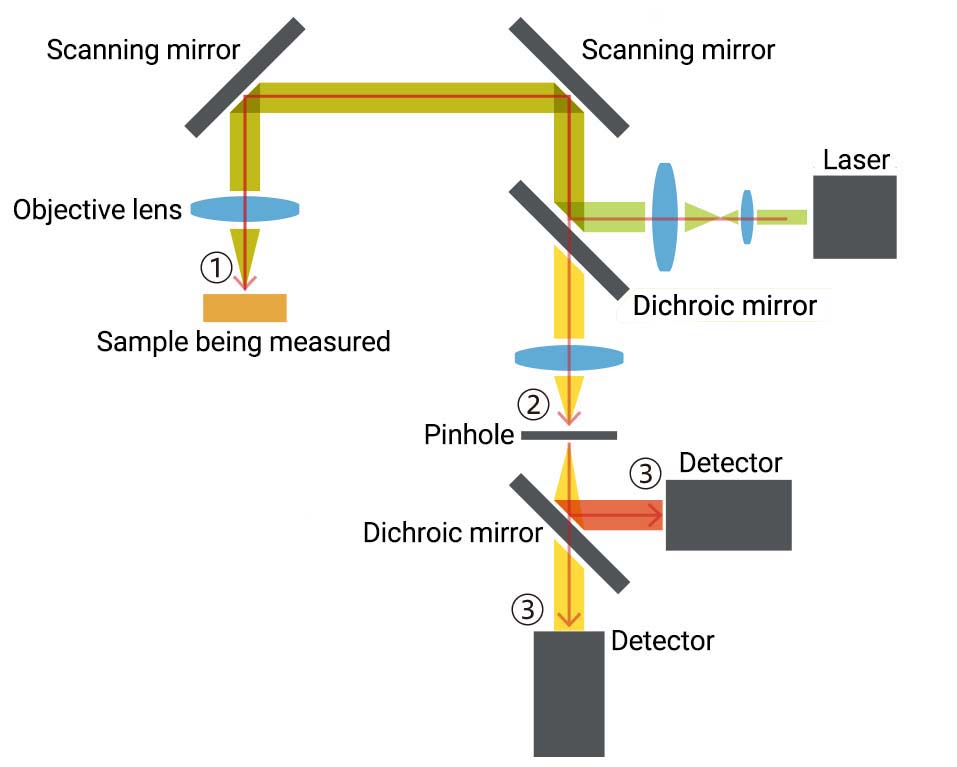
Principle of laser confocal microscope
There are various types of Laser scanning microscopes depending on the application and detector configuration, so their internal structure and operating principle differ from each other.
Here, we introduce the principle of a typical laser confocal microscope as an example.
- Laser light strikes a target object (sample) via scanning mirrors.
- Fluorescence emitted from the sample is directed to a pinhole.
- The scattered light is reduced by the pinhole placed at the focus point and is measured by the detector.
Role of the pinhole in fluorescence measurement
The method of scanning the laser light and the configuration of the optical system differ depending on each type of microscope. One distinct feature of laser confocal microscopes is the pinhole placed at the focus point. The pinhole eliminates light unnecessary for measurement so that only fluorescence very close to the focal plane of the sample can be detected. Due to this structure, laser confocal microscopes allow measurements with excellent optical resolution and resolving power in the depth direction.
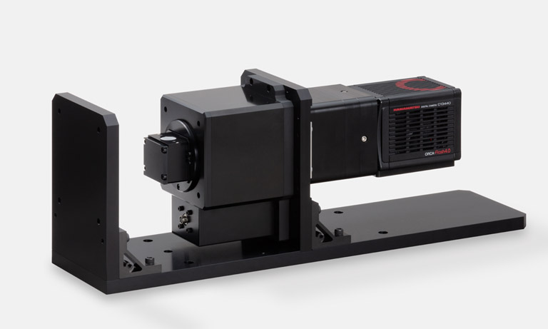
Development of key devices for Laser scanning microscopes
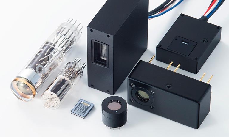
Hamamatsu Photonics develops and manufactures a large number of key devices that are essential for Laser scanning microscope operation and performance.
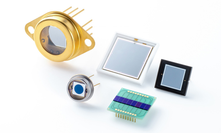
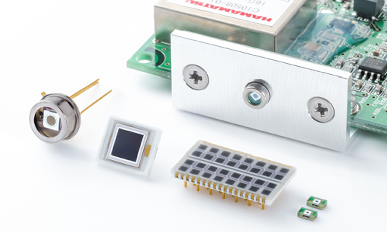
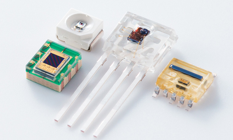
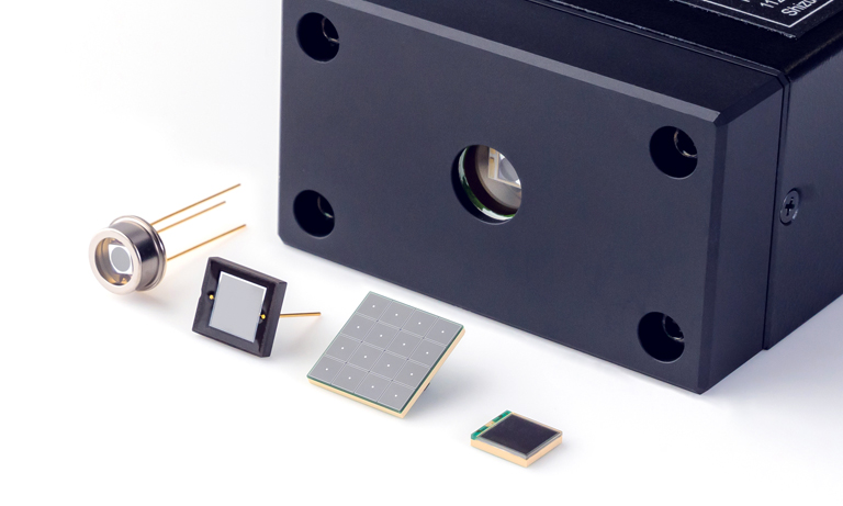
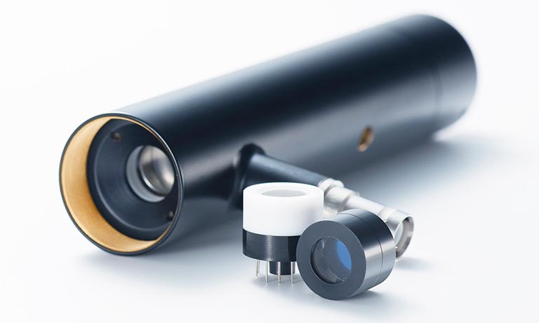
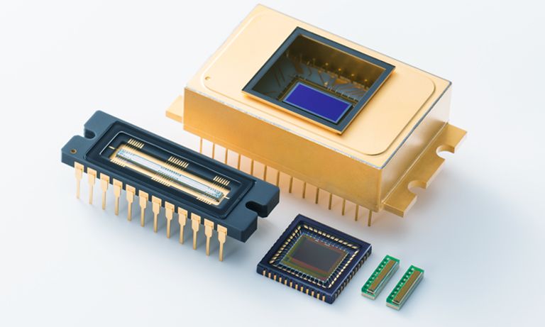
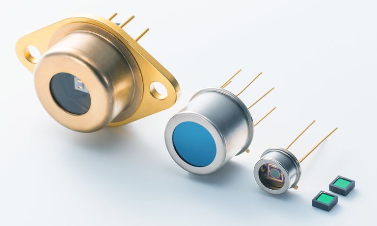
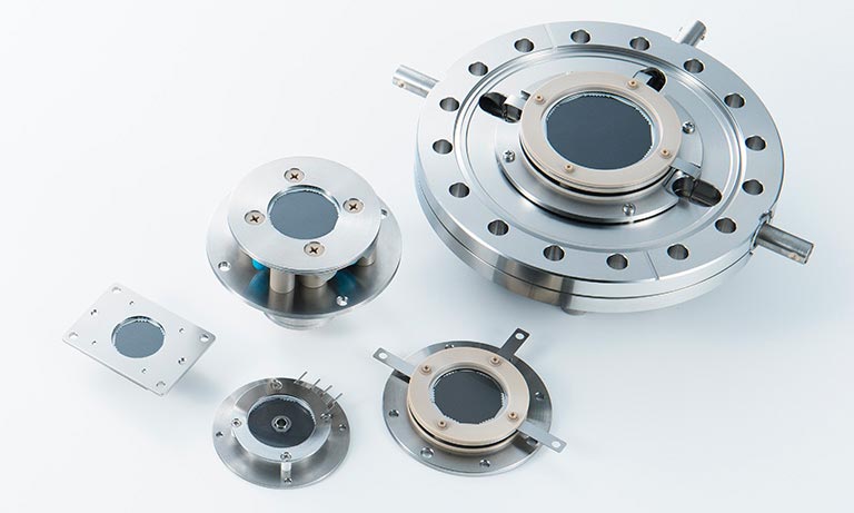
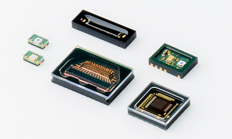
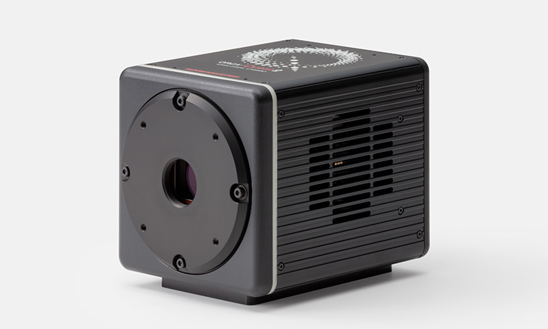
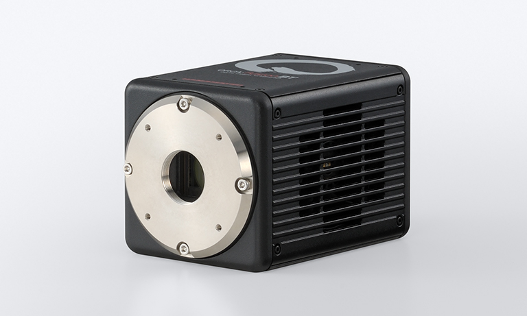
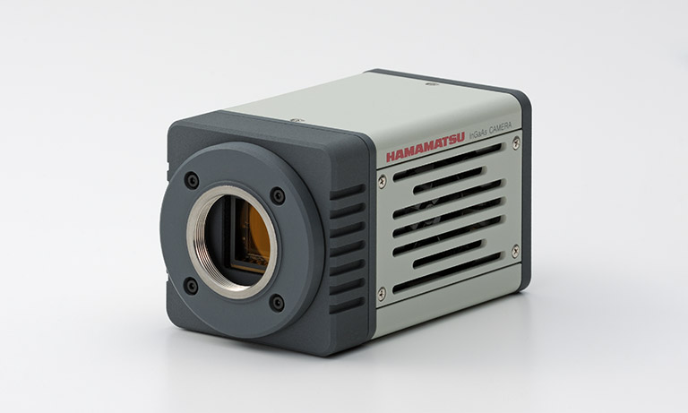
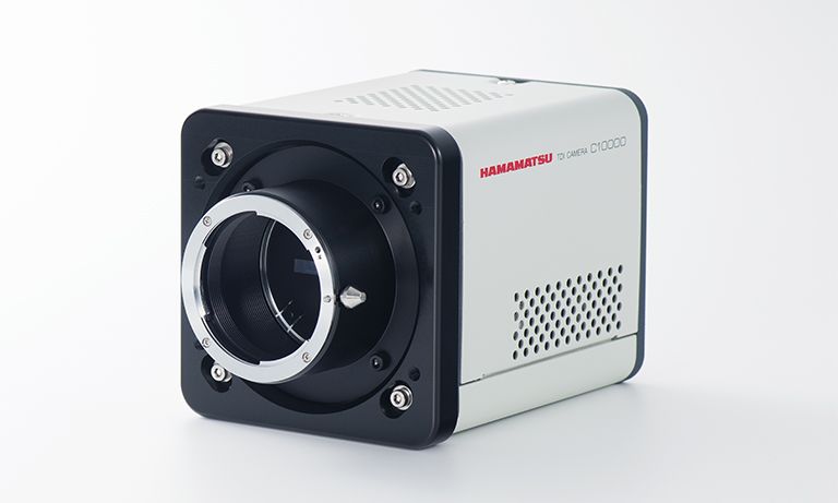
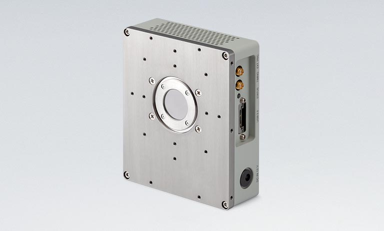
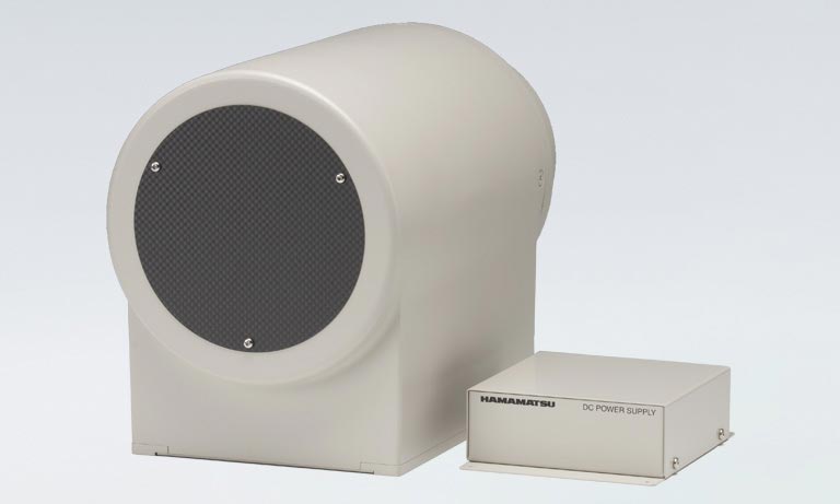
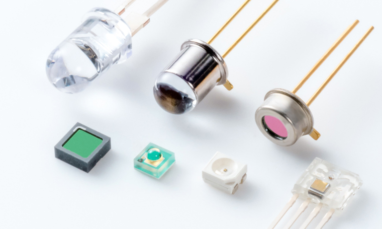
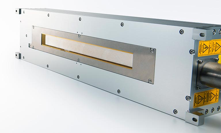
Photodetectors
Photodetectors are the core of Laser scanning microscopes and are directly linked to the quality of the acquired image. Among photodetectors, photomultiplier tubes (PMT) with excellent sensitivity and noise characteristics and multi-pixel photon counters (MPPC) which are often called silicon photomultipliers or Si-PM are widely used to acquire microscopic images with ever higher quality.
Features of photodetectors
- High sensitivity: Boosts image quality and resolution of acquired images.
- Low noise: Boosts image quality of acquired images.
- Wide dynamic range: Boosts image quality of acquired images.

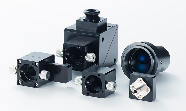
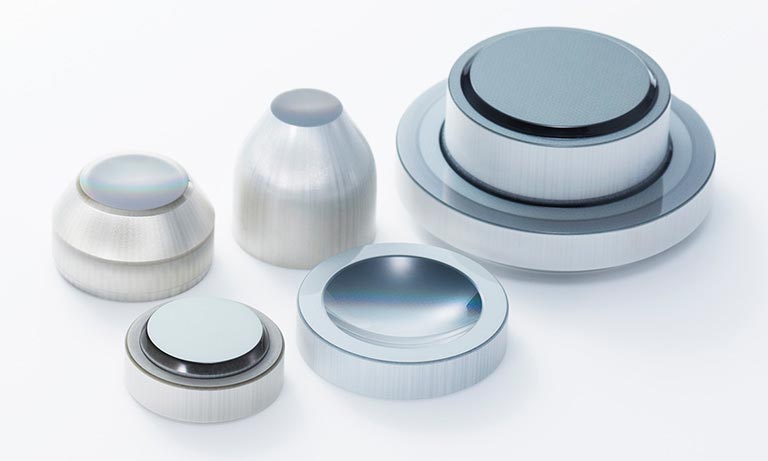
Optical components (mirrors)
Hamamatsu Photonics develops and manufactures MEMS (Micro-Electro-Mechanical Systems) mirrors that offer many advantages over Galvano mirrors which have been widely used as scanning mirrors. These advantages include a subminiature size, low cost and low power consumption. Two-dimensional scanning is now possible with a single MEMS mirror device, which will make devices easy to downsize and drastically speed up equipment operation.
Features of optical components (mirrors)
- Miniature size: Helps shrink the equipment size.
- High-speed operation: Trims the image acquisition time.
- Two-dimensional scanning: Does two-dimensional scanning with a single device.
Selecting detectors
Detectors for Laser scanning microscopes must exhibit excellent characteristics in sensitivity and noise. This is important because the amount of light incident on the detector is limited due to the basic measurement principle of Laser scanning microscopes.
Ex. 1 Laser confocal microscope: The pinhole reduces the amount of light incident on the detector.
Ex. 2 Multiphoton microscope: The probability that the multiphoton absorption phenomenon will occur is extremely low so the amount of light is limited.
There are various types of detectors having different functions, so it is essential to select the optimal detector that best matches the microscope specifications.
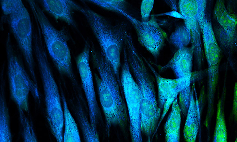
The photos show images of the same sample observed using different PMTs serving as the detector for a Laser scanning microscope.
Compared to the left image acquired using a multialkali photocathode PMT, the right image acquired using a GaAsP crystal photocathode PMT shows clear and distinct contrast. As seen from this example, even when using a high sensitivity detector like a PMT, the acquired image will greatly differ depending on its photocathode type.
Hamamatsu Photonics offers a broad lineup of detectors that allow you to select the optimal detector to meet your measurement criteria and conditions such as the light level, wavelength, sample thickness, scanning speed, and cooling method.
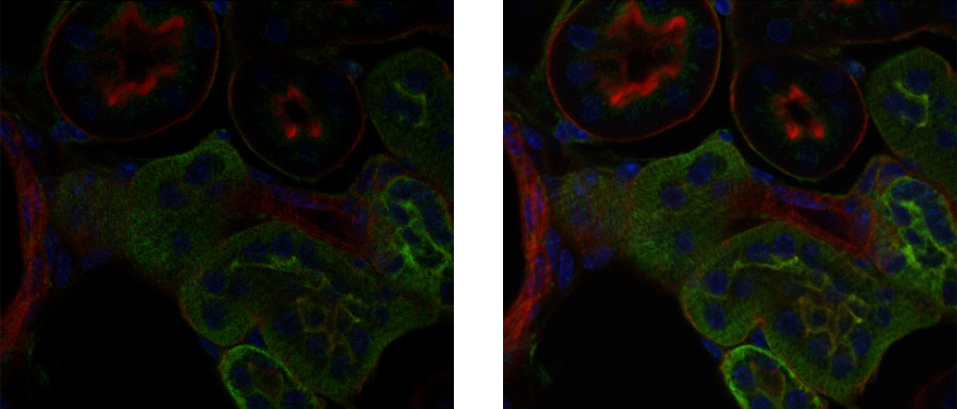
Left: Image acquired using multialkali photocathode PMT
Right: Image acquired using GaAsP photocathode PMT
※Sample: Mouse cells
Related Document
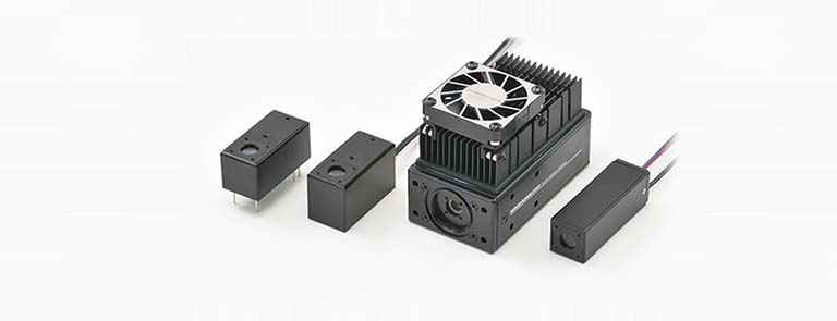
A series of highly sensitive photomultiplier tube modules with a crystalline photocathode are collectively introduced.
Quantum efficiency color chart
Hamamatsu Photonics offers a broad lineup of detectors, providing the optimum detector based on many criteria such as light intensity, measurement wavelength, sample thickness, and scanning speed. Among them, high-sensitivity photomultiplier tubes contribute greatly to the image quality of laser microscopes.
By using crystal materials such as GaAsP and GaAs for the parts that convert light incident on the photomultiplier tube into electrons, higher quantum efficiency is achieved compared to types using photocathodes made of ordinary alkali materials.
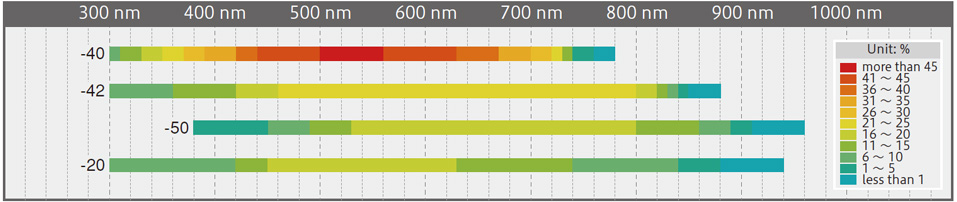
The spectral response range is defined differently depending on the type of photocathode. For details, see P. 04 of the catalog.
Measurement techniques and recommended products
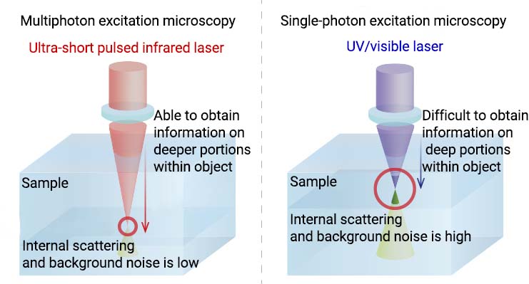
Multiphoton microscopy
Multiphoton microscopy is a technique for observing fluorescence in the UV to visible region that occurs when fluorescent molecules are excited by simultaneously absorbing two photons.
The wavelength of the excitation light in common fluorescence measurement is shorter than the fluorescence wavelength. However, multiphoton spectroscopy uses excitation light in the near-infrared region, which has a wavelength longer than the fluorescence wavelength. Since near-infrared light passes more easily through an object than visible light does, it can provide information on deep portions within an object and also reduces effects from scattering and background noise inside the object. Furthermore, the energy of near-infrared light is lower than that of visible and UV light, thus minimizing damage to cells.
■ How it differs from single-photon excitation microscopy
Imaging example: Observation of deep mouse brain
・Imaging depth: 500 µm from brain surface
・Imaging range: 3 mm × 3 mm
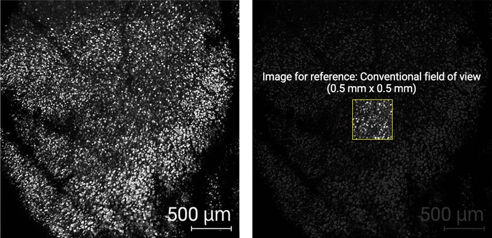
The image was obtained by detecting signals with a high signal-to-noise ratio using a high sensitivity photomultiplier tube (equivalent to Hamamatsu H15460-40). This photomultiplier tube also has a wide photosensitive area so that observation of deep portions, which is a unique feature of multiphoton microscopy, is possible in a wide field of view.
Image courtesy:RIKEN Center for Brain Science(CBS) Masanori Murayama (Ph.D.)
Recommended products for multiphoton microscopy
| Products category | Image | Products name | Features |
|---|---|---|---|
| Detectors | 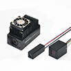 |
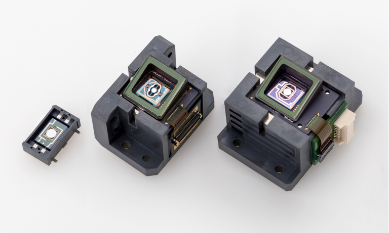
Photomultiplier tube module H15460-40 | Large photosensitive area: 14 mm sq. |
| Photomultiplier tube module H16722-40 | Built-in thermoelectric cooler: Minimizes thermal noise | ||
 |
MPPC modules C13852 series | Compact and lightweight unit with visible light sensitivity | |
| MPPC modules C14456 series | Compact and lightweight unit with visible to near-infrared sensitivity | ||
| Optical components | 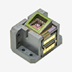 |
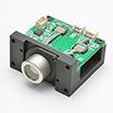
MEMS mirror S13989-01H | Two-dimensional scanning by reflection of laser light |
 |
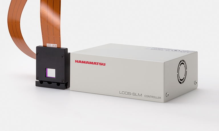
LCOS-SLM X15213 series | Phase Control (Wevefront control) for aberration correction |
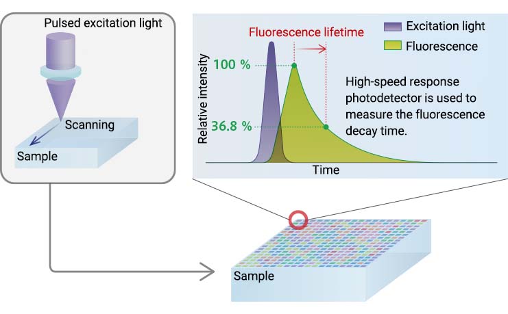
Fluorescence lifetime imaging microscopy (FLIM)
FLIM is a technique for measuring the decay time (lifetime) of fluorescence emitted from an object.
FLIM measures the fluorescence lifetime unique to each fluorescent molecule by time-resolved measurement, and in this way gives users more information than is possible just from conventional fluorescence measurement. The detectors used for FLIM are required to have both high-speed response and high sensitivity since they must measure small changes in fluorescence intensity occurring within an extremely short time.
Recommended products for FLIM
| Products category | Image | Products name | Features |
|---|---|---|---|
| Detectors | 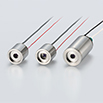 |
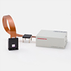
HPD | High-speed response High time resolution |
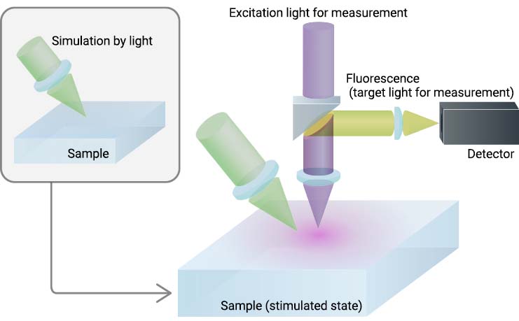
Photostimulation measurement
This technique stimulates a sample with light and observes its reaction in real time. It is mainly used for measuring biological samples. Since multiple lasers for excitation and stimulation are used in photostimulation measurement, it is essential to create an optical design that accurately detects the measurement light at the required timing.
Photomultiplier tube modules with a gate function are ideal as devices that mount in a microscope utilizing photostimulation since their gate operation can be electrically controlled to match the timing of light detection.
Moreover, by splitting a laser beam into multiple beams with an optical system using LCOS-SLM wavefront shaping technology, multiple points can be simultaneously stimulated by light with a single laser unit.
Recommended products for photostimulation measurements
| Products category | Image | Products name | Features |
|---|---|---|---|
| Detectors |  |
Photomultiplier tube module H12056-40 | Compact unit integrated with a gate circuit Maximum output signal current: 40 µA |
| Photomultiplier tube module H11706-40 | Compact unit integrated with a gate circuit Maximum output signal current: 2 µA |
||
|
MPPC modules C13852 series | Compact and lightweight unit with visible light sensitivity | |
| MPPC modules C14456 series | Compact and lightweight unit with visible to near-infrared sensitivity | ||
| Optical components | |
MEMS mirror S13989-01H | Two-dimensional scanning by reflection of laser light |
|
LCOS-SLM X15213 series | Phase control (wavefront control) for generating multiple point |
Multicolor measurements
This technique is recently the focus of a huge amount of attention due to increasing needs for measuring longer wavelength fluorescence emitted from fluorescent proteins. Multicolor measurements capture information over a broad range of the spectrum from visible to near infrared, making this a promising way to reveal phenomena that have not been seen before.
Photodetectors with a wide spectral response range that is also highly sensitive in the near-infrared region are effective.

Recommended products for multicolor measurements
| Products category | Image | Products name | Features |
|---|---|---|---|
| Detectors |  |
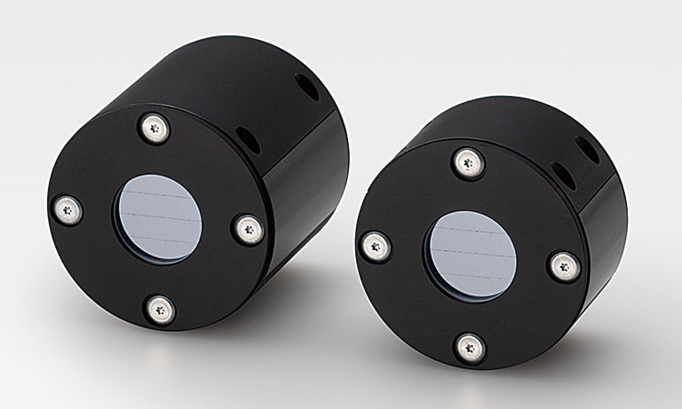
Photomultiplier tube R13456 | High sensitivity in the near-infrared region |
| Photomultiplier tube R10699 | Wide spectral response range from visible to near-infrared | ||
|
MPPC modules C13852 series | Compact and lightweight unit with visible light sensitivity | |
| MPPC modules C14456 series | Compact and lightweight unit with visible to near-infrared sensitivity |
- Confirmation
-
It looks like you're in the . If this is not your location, please select the correct region or country below.
You're headed to Hamamatsu Photonics website for GB (English). If you want to view an other country's site, the optimized information will be provided by selecting options below.
In order to use this website comfortably, we use cookies. For cookie details please see our cookie policy.
- Cookie Policy
-
This website or its third-party tools use cookies, which are necessary to its functioning and required to achieve the purposes illustrated in this cookie policy. By closing the cookie warning banner, scrolling the page, clicking a link or continuing to browse otherwise, you agree to the use of cookies.
Hamamatsu uses cookies in order to enhance your experience on our website and ensure that our website functions.
You can visit this page at any time to learn more about cookies, get the most up to date information on how we use cookies and manage your cookie settings. We will not use cookies for any purpose other than the ones stated, but please note that we reserve the right to update our cookies.
1. What are cookies?
For modern websites to work according to visitor’s expectations, they need to collect certain basic information about visitors. To do this, a site will create small text files which are placed on visitor’s devices (computer or mobile) - these files are known as cookies when you access a website. Cookies are used in order to make websites function and work efficiently. Cookies are uniquely assigned to each visitor and can only be read by a web server in the domain that issued the cookie to the visitor. Cookies cannot be used to run programs or deliver viruses to a visitor’s device.
Cookies do various jobs which make the visitor’s experience of the internet much smoother and more interactive. For instance, cookies are used to remember the visitor’s preferences on sites they visit often, to remember language preference and to help navigate between pages more efficiently. Much, though not all, of the data collected is anonymous, though some of it is designed to detect browsing patterns and approximate geographical location to improve the visitor experience.
Certain type of cookies may require the data subject’s consent before storing them on the computer.
2. What are the different types of cookies?
This website uses two types of cookies:
- First party cookies. For our website, the first party cookies are controlled and maintained by Hamamatsu. No other parties have access to these cookies.
- Third party cookies. These cookies are implemented by organizations outside Hamamatsu. We do not have access to the data in these cookies, but we use these cookies to improve the overall website experience.
3. How do we use cookies?
This website uses cookies for following purposes:
- Certain cookies are necessary for our website to function. These are strictly necessary cookies and are required to enable website access, support navigation or provide relevant content. These cookies direct you to the correct region or country, and support security and ecommerce. Strictly necessary cookies also enforce your privacy preferences. Without these strictly necessary cookies, much of our website will not function.
- Analytics cookies are used to track website usage. This data enables us to improve our website usability, performance and website administration. In our analytics cookies, we do not store any personal identifying information.
- Functionality cookies. These are used to recognize you when you return to our website. This enables us to personalize our content for you, greet you by name and remember your preferences (for example, your choice of language or region).
- These cookies record your visit to our website, the pages you have visited and the links you have followed. We will use this information to make our website and the advertising displayed on it more relevant to your interests. We may also share this information with third parties for this purpose.
Cookies help us help you. Through the use of cookies, we learn what is important to our visitors and we develop and enhance website content and functionality to support your experience. Much of our website can be accessed if cookies are disabled, however certain website functions may not work. And, we believe your current and future visits will be enhanced if cookies are enabled.
4. Which cookies do we use?
There are two ways to manage cookie preferences.
- You can set your cookie preferences on your device or in your browser.
- You can set your cookie preferences at the website level.
If you don’t want to receive cookies, you can modify your browser so that it notifies you when cookies are sent to it or you can refuse cookies altogether. You can also delete cookies that have already been set.
If you wish to restrict or block web browser cookies which are set on your device then you can do this through your browser settings; the Help function within your browser should tell you how. Alternatively, you may wish to visit www.aboutcookies.org, which contains comprehensive information on how to do this on a wide variety of desktop browsers.
5. What are Internet tags and how do we use them with cookies?
Occasionally, we may use internet tags (also known as action tags, single-pixel GIFs, clear GIFs, invisible GIFs and 1-by-1 GIFs) at this site and may deploy these tags/cookies through a third-party advertising partner or a web analytical service partner which may be located and store the respective information (including your IP-address) in a foreign country. These tags/cookies are placed on both online advertisements that bring users to this site and on different pages of this site. We use this technology to measure the visitors' responses to our sites and the effectiveness of our advertising campaigns (including how many times a page is opened and which information is consulted) as well as to evaluate your use of this website. The third-party partner or the web analytical service partner may be able to collect data about visitors to our and other sites because of these internet tags/cookies, may compose reports regarding the website’s activity for us and may provide further services which are related to the use of the website and the internet. They may provide such information to other parties if there is a legal requirement that they do so, or if they hire the other parties to process information on their behalf.
If you would like more information about web tags and cookies associated with on-line advertising or to opt-out of third-party collection of this information, please visit the Network Advertising Initiative website http://www.networkadvertising.org.
6. Analytics and Advertisement Cookies
We use third-party cookies (such as Google Analytics) to track visitors on our website, to get reports about how visitors use the website and to inform, optimize and serve ads based on someone's past visits to our website.
You may opt-out of Google Analytics cookies by the websites provided by Google:
https://tools.google.com/dlpage/gaoptout?hl=en
As provided in this Privacy Policy (Article 5), you can learn more about opt-out cookies by the website provided by Network Advertising Initiative:
http://www.networkadvertising.org
We inform you that in such case you will not be able to wholly use all functions of our website.
Close