
Products










We are actively taking measures to improve product quality levels.
Applications















Why Hamamatsu?
Resources





Support




Our company







Investors








United Kingdom (EN)
Select your region or country.

Photonics brings new light to brain monitoring: Advancing non-invasive monitoring through optical sensing
Shining a light on the brain: Advancing non-invasive monitoring through optical sensing
Understanding the brain in real time remains one of the most complex challenges in modern medicine. Monitoring cerebral oxygenation can be critical in clinical environments such as neurosurgery, intensive care, and emergency medicine, helping to guide interventions and improve patient outcomes. However, many established techniques have clear limitations; invasive approaches – such as intracranial pressure (ICP) monitoring – carry inherent risks and are not suitable for all patients, while imaging modalities such as MRI and CT require comparatively long acquisition times, preventing continuous real-time monitoring, as well as being immobile and resource-intensive.
Healthcare is moving toward more continuous, patient-centric monitoring, driven by the need to detect physiological changes earlier, tailor interventions to individual patients, and reduce reliance on single time-point measurements. This has created growing demand for technologies that can deliver real-time insights non-invasively. Photonics is playing an increasingly important role in meeting this need, with emerging approaches such as functional near-infrared spectroscopy (fNIRS) gaining attention, thanks to the combination of accessibility with meaningful physiological measurements they offer.
The optical principle
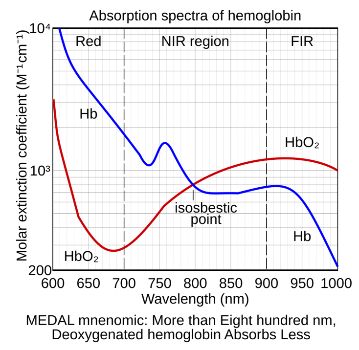
The foundations of fNIRS lie in the interaction between near-infrared light and biological tissues. Light in the wavelength range of approximately 650 to 1100 nm can penetrate biological tissue, providing a non-invasive optical window into underlying physiological processes.1 fNIRS uses this principle to measure changes in hemoglobin concentration within the brain. By emitting multiple wavelengths of near-infrared light and detecting the returning signal, it becomes possible to distinguish between oxygenated and deoxygenated hemoglobin, reflecting regional changes in neuronal activation.1,2 However, in practice, only a small fraction of the emitted light reaches the detector, so the resulting signal is weak and highly sensitive to noise, making measurements technically demanding.
Newer optical approaches are also expanding the scope of non-invasive brain monitoring. Techniques such as time-domain near-infrared spectroscopy (TD-NIRS) and diffuse correlation spectroscopy (DCS) can provide additional insights into cerebral perfusion, blood flow, and hemodynamic function, broadening the physiological information available through optical neuromonitoring.3
Engineering challenges in optical brain monitoring
Realizing the potential of fNIRS for brain monitoring relies on overcoming a complex set of engineering constraints. One of the primary challenges is signal attenuation and scattering. As near-infrared light travels through the scalp, skull, and brain tissue, it is both absorbed and diffusely scattered, resulting in a substantial loss of signal intensity. By the time light reaches the detector, only a small fraction of the original photons remain, making the system highly sensitive to noise and requiring detectors with high-sensitivity and low-noise characteristics. Closely related to this is the trade-off between measurement depth and signal strength, where the separation between the light source and detector determines how deeply the light penetrates into the tissue. Larger separations allow interrogation of deeper brain regions, but reduce the intensity of the detected signal, while shorter separations provide stronger signals that are more heavily influenced by superficial tissues. Balancing this is a key aspect of system design.
Another challenge arises from the mixed nature of the detected signal. Measurements often include contributions from extracerebral tissues – such as the scalp and skull – in addition to the brain itself. This issue has been recognized since early clinical investigations, which highlighted extracranial signal contamination as a key limitation, while also demonstrating that near-infrared measurements can still reflect intracranial hemodynamic changes under controlled conditions. Isolating the cerebral component therefore requires careful system design and signal processing, particularly in applications where accuracy is critical.
Real-world variability further complicates measurement. Factors such as skin tone, hair, and movement can affect optical coupling and signal quality. Furthermore, motion – such as during ambulance rides or long-term monitoring – can introduce significant noise. Ambient light interference must also be managed to ensure reliable operation outside of controlled environments. Finally, the physical interface between the system and the patient plays a crucial role. Maintaining consistent contact between sensors and the skin is essential for repeatable measurements. This places constraints on sensor design, attachment mechanisms, and overall system ergonomics, particularly for wearable or long-duration applications. These challenges put significant demands on both the optical components and the overall system architecture.
From components to complete systems
Specialized optical components designed to operate in the near-infrared spectrum are at the heart of fNIRS systems. Light sources typically include lasers and LEDs capable of delivering stable outputs at specific wavelengths, while detectors such as silicon and InGaAs photodiodes, avalanche photodiodes (APDs), and silicon photomultipliers (SiPMs or MPPCs) are used to capture the returning signal.
System performance depends on several key parameters. Wavelength selection, typically in the 650 to 1100 nm range, is critical for differentiating between oxygenated and deoxygenated hemoglobin, as well as allowing depth selectivity. Detector sensitivity and noise characteristics directly influence the ability to capture weak signals, while dynamic range determines how well the system can accommodate varying signal intensities.
Modern systems often incorporate multiple source–detector pairs arranged across the scalp, enabling spatial mapping of brain activity. While this increases the amount of information that can be captured, it also adds complexity in terms of synchronization, data acquisition, and signal processing. As a result, successful implementation depends not only on individual component performance, but on how these elements are integrated into a complete system. Customization is frequently required to meet the needs of specific applications, whether prioritizing depth sensitivity, spatial resolution, portability, or user comfort. Achieving the right balance between these factors is central to delivering meaningful and reliable measurements. When these challenges are addressed, fNIRS can be applied across a growing range of clinical and research settings.
Clinical and research applications of fNIRS
One of the main strengths of fNIRS is its flexibility. Unlike many traditional neuroimaging techniques, it can be deployed in a wide range of environments, from controlled laboratory studies to bedside monitoring. In clinical settings, fNIRS is increasingly being explored for monitoring cerebral oxygenation during surgery and in critical care. Continuous measurement of oxygenation in these settings has the potential to provide early warning of physiological deterioration, particularly in cases such as traumatic brain injury or compromised cerebral perfusion. Emerging research suggests that features derived from optical signals may correlate with changes in intracranial pressure, highlighting the potential for non-invasive alternatives to current monitoring techniques. Earlier clinical studies have also shown that fNIRS can track cerebral hemodynamic changes in surgical settings, reinforcing the physiological relevance of these measurements.4
Beyond acute care, fNIRS has also shown benefits in neuroscience research, particularly in populations where other methods are difficult to apply. Its non-invasive nature and tolerance to movement compared to techniques like MRI make it especially suitable for use in infants and children. Because it does not rely on behavioral responses, it enables investigation of brain function even in very early developmental stages. This has opened new avenues for studying cognitive development, social processing, and neurological conditions such as autism spectrum disorders. Researchers can examine how the brain responds to stimuli, how functional networks develop over time, and how these processes may differ in atypical development.5
In addition to developmental studies, fNIRS is being used in functional brain mapping. By measuring hemodynamic responses during tasks, it allows researchers to explore the relationship between brain activity and behavior. This capability forms the foundation for more advanced imaging approaches, including higher resolution and three-dimensional brain mapping techniques.1,6 However, translating these capabilities into robust, clinically-reliable systems requires careful consideration of the underlying technical challenges.
Beyond fNIRS
fNIRS focuses on hemodynamic responses, making it just part of a broader landscape of brain monitoring technologies. Diffuse Optical Tomography (DOT) extends the principles of fNIRS by using multiple sources and detectors to reconstruct three-dimensional maps of brain activity. This approach provides improved spatial resolution, and is widely used in research settings to study functional connectivity and brain organization.6 In contrast, magnetoencephalography (MEG) measures the magnetic fields generated by neuronal electrical activity. This allows for high temporal resolution and direct measurement of neural signals. However, conventional MEG systems are large, expensive, and require specialized environments. Recent developments in optically pumped magnetometers (OPMs) are beginning to address these limitations. By enabling more compact and flexible MEG systems, they offer the potential for improved patient comfort and broader accessibility. Together, these approaches highlight the expanding role of photonics in enabling new modalities for brain monitoring.
Enabling continuous, non-invasive brain monitoring
Advances in both hardware and data analysis are beginning to unlock new possibilities for fNIRS. Research has shown that features extracted from optical signals can differentiate between physiological states associated with changes in intracranial pressure, achieving promising levels of sensitivity in early-stage studies. While these approaches are still under development, they point toward a future in which continuous, non-invasive brain monitoring could complement existing techniques. Improvements in sensor design and detector performance are helping to improve signal quality and reliability, while there is a clear trend toward more portable and wearable systems.
This is enabling monitoring to move beyond traditional clinical environments, with potential applications in bedside, ambulatory, and remote settings. Integration with other modalities, such as electroencephalography (EEG), also offers opportunities to combine complementary information and improve overall insights into brain activity.
Illuminating the future of brain monitoring
The demand for accessible, real-time brain monitoring continues to grow, driven by the need for earlier intervention and more personalised care. Photonics-based technologies are playing an increasingly important role in meeting this need, enabling new approaches that combine non-invasiveness with meaningful physiological insights. fNIRS illustrates both the progress that has been made and the challenges that remain. While its portability and versatility open up new possibilities, achieving consistent, high-quality measurements in real-world settings requires continued innovation in both system design and application development. Progress in this field will depend on close collaboration between researchers, clinicians, and photonics experts. By combining application expertise with advances in optical technologies, it is possible to move closer to reliable, non-invasive brain monitoring that can be applied at scale. Companies that develop photonic components and systems will continue to play an important role in translating research advances into clinical practice.
Related products
References
1) Chen WL, Wagner J, Heugel N, Sugar J, Lee YW, Conant L, Malloy M, Heffernan J, Quirk B, Zinos A, Beardsley SA, Prost R, Whelan HT. Functional Near-Infrared Spectroscopy and Its Clinical Application in the Field of Neuroscience: Advances and Future Directions. Front Neurosci. 2020 Jul 9;14:724. doi: 10.3389/fnins.2020.00724.
2) Pinti P, Tachtsidis I, Hamilton A, Hirsch J, Aichelburg C, Gilbert S, Burgess PW. The present and future use of functional near-infrared spectroscopy (fNIRS) for cognitive neuroscience. Ann N Y Acad Sci. 2020 Mar;1464(1):5-29. doi: 10.1111/nyas.13948.
3) Carp S, Tamborini D, Mazumder D, Wu KC, Robinson M, Stephens K, Shatrovoy O, Lue N, Ozana N, Blackwell M, Franceschini MA. Diffuse correlation spectroscopy measurements of blood flow using 1064 nm light. J Biomed Opt. 2020 Sep;25(9):097003. doi: 10.1117/1.JBO.25.9.097003.
4) Al-Rawi PG, Smielewski P, Kirkpatrick PJ. Stroke. 2001 Nov;32(11):2492-500. doi: 10.1161/hs1101.098356.
5) Lloyd-Fox, S. Functional near-infrared spectroscopy. The Cambridge Encyclopedia of Child Development 129–136. Cambridge University Press. 2017. doi:10.1017/9781316216491.022.
6) Fogarty M, Rafferty SM, Markow ZE, O'Sullivan AC, Svoboda CF, George T, King K, Wilhelm D, Tripathy K, Mugler EM, Naufel S, Yin A, Trobaugh JW, Eggebrecht AT, Richter EJ, Culver JP. Functional brain mapping using whole-head very high-density diffuse optical tomography. Imaging Neurosci (Camb). 2025 Jun 20;3:IMAG.a.54. doi: 10.1162/IMAG.a.54.
- Confirmation
-
It looks like you're in the . If this is not your location, please select the correct region or country below.
You're headed to Hamamatsu Photonics website for GB (English). If you want to view an other country's site, the optimized information will be provided by selecting options below.
In order to use this website comfortably, we use cookies. For cookie details please see our cookie policy.
- Cookie Policy
-
This website or its third-party tools use cookies, which are necessary to its functioning and required to achieve the purposes illustrated in this cookie policy. By closing the cookie warning banner, scrolling the page, clicking a link or continuing to browse otherwise, you agree to the use of cookies.
Hamamatsu uses cookies in order to enhance your experience on our website and ensure that our website functions.
You can visit this page at any time to learn more about cookies, get the most up to date information on how we use cookies and manage your cookie settings. We will not use cookies for any purpose other than the ones stated, but please note that we reserve the right to update our cookies.
1. What are cookies?
For modern websites to work according to visitor’s expectations, they need to collect certain basic information about visitors. To do this, a site will create small text files which are placed on visitor’s devices (computer or mobile) - these files are known as cookies when you access a website. Cookies are used in order to make websites function and work efficiently. Cookies are uniquely assigned to each visitor and can only be read by a web server in the domain that issued the cookie to the visitor. Cookies cannot be used to run programs or deliver viruses to a visitor’s device.
Cookies do various jobs which make the visitor’s experience of the internet much smoother and more interactive. For instance, cookies are used to remember the visitor’s preferences on sites they visit often, to remember language preference and to help navigate between pages more efficiently. Much, though not all, of the data collected is anonymous, though some of it is designed to detect browsing patterns and approximate geographical location to improve the visitor experience.
Certain type of cookies may require the data subject’s consent before storing them on the computer.
2. What are the different types of cookies?
This website uses two types of cookies:
- First party cookies. For our website, the first party cookies are controlled and maintained by Hamamatsu. No other parties have access to these cookies.
- Third party cookies. These cookies are implemented by organizations outside Hamamatsu. We do not have access to the data in these cookies, but we use these cookies to improve the overall website experience.
3. How do we use cookies?
This website uses cookies for following purposes:
- Certain cookies are necessary for our website to function. These are strictly necessary cookies and are required to enable website access, support navigation or provide relevant content. These cookies direct you to the correct region or country, and support security and ecommerce. Strictly necessary cookies also enforce your privacy preferences. Without these strictly necessary cookies, much of our website will not function.
- Analytics cookies are used to track website usage. This data enables us to improve our website usability, performance and website administration. In our analytics cookies, we do not store any personal identifying information.
- Functionality cookies. These are used to recognize you when you return to our website. This enables us to personalize our content for you, greet you by name and remember your preferences (for example, your choice of language or region).
- These cookies record your visit to our website, the pages you have visited and the links you have followed. We will use this information to make our website and the advertising displayed on it more relevant to your interests. We may also share this information with third parties for this purpose.
Cookies help us help you. Through the use of cookies, we learn what is important to our visitors and we develop and enhance website content and functionality to support your experience. Much of our website can be accessed if cookies are disabled, however certain website functions may not work. And, we believe your current and future visits will be enhanced if cookies are enabled.
4. Which cookies do we use?
There are two ways to manage cookie preferences.
- You can set your cookie preferences on your device or in your browser.
- You can set your cookie preferences at the website level.
If you don’t want to receive cookies, you can modify your browser so that it notifies you when cookies are sent to it or you can refuse cookies altogether. You can also delete cookies that have already been set.
If you wish to restrict or block web browser cookies which are set on your device then you can do this through your browser settings; the Help function within your browser should tell you how. Alternatively, you may wish to visit www.aboutcookies.org, which contains comprehensive information on how to do this on a wide variety of desktop browsers.
5. What are Internet tags and how do we use them with cookies?
Occasionally, we may use internet tags (also known as action tags, single-pixel GIFs, clear GIFs, invisible GIFs and 1-by-1 GIFs) at this site and may deploy these tags/cookies through a third-party advertising partner or a web analytical service partner which may be located and store the respective information (including your IP-address) in a foreign country. These tags/cookies are placed on both online advertisements that bring users to this site and on different pages of this site. We use this technology to measure the visitors' responses to our sites and the effectiveness of our advertising campaigns (including how many times a page is opened and which information is consulted) as well as to evaluate your use of this website. The third-party partner or the web analytical service partner may be able to collect data about visitors to our and other sites because of these internet tags/cookies, may compose reports regarding the website’s activity for us and may provide further services which are related to the use of the website and the internet. They may provide such information to other parties if there is a legal requirement that they do so, or if they hire the other parties to process information on their behalf.
If you would like more information about web tags and cookies associated with on-line advertising or to opt-out of third-party collection of this information, please visit the Network Advertising Initiative website http://www.networkadvertising.org.
6. Analytics and Advertisement Cookies
We use third-party cookies (such as Google Analytics) to track visitors on our website, to get reports about how visitors use the website and to inform, optimize and serve ads based on someone's past visits to our website.
You may opt-out of Google Analytics cookies by the websites provided by Google:
https://tools.google.com/dlpage/gaoptout?hl=en
As provided in this Privacy Policy (Article 5), you can learn more about opt-out cookies by the website provided by Network Advertising Initiative:
http://www.networkadvertising.org
We inform you that in such case you will not be able to wholly use all functions of our website.
Close