![]()
Products


We are actively taking measures to improve product quality levels.
Applications


Why Hamamatsu?
Resources


Support


Our company


Investors


United Kingdom (EN)
Select your region or country.

How Flow Cytometry Works: Optics, Lasers and Photon Detectors
Flow cytometry enables fast, quantitative insights at single-cell level for research and clinical use. It is widely used to identify immune cell subsets, detect rare cells in blood, and monitor cell health in bioprocesses. Reliable results are therefore paramount. This is why the instrument’s optical system matters, especially the lasers, filters, and detectors that generate and capture the signal. In this article, we follow the path from sample to signal and how optical design and detector choice, including PMTs, APDs, and SiPMs, influence sensitivity, linearity, and multiplexing.
Why flow cytometry— and why reliability is hard
Flow cytometry can deliver fast results because it interrogates cells as they move through an optical system, but the same speed makes the measurement chain unforgiving. Weak fluorescence signals, spectral overlap, optical losses, and detector noise can all affect how clearly cell populations are identified and quantified. This is why the optical chain, from laser excitation and spectral separation to photon detection must be designed for stable, efficient signal capture.
How flow cytometry generates signals
In flow cytometry, cells suspended in a fluid stream are hydrodynamically aligned in single file and pass through one or more focused laser beams for optical interrogation. Detectors capture scatter and fluorescence to quantify cell properties.
The interaction of incident light with cells generates two main optical signals: light scattering and fluorescence.
Light scattering measurements
- Forward Scatter – primarily correlates with cell or particle size
- Side Scatter – reflects internal structural complexity or granularity
Fluorescence detection
Fluorescence signals originate from fluorophore-labeled antibodies, chemical dyes, or genetically encoded reporters. These optical signals are collected by photodetectors, converted into electrical pulses, and digitally processed to generate quantitative cellular parameters used for multiparametric single-cell analysis.
The optical chain in flow cytometry
Reliable measurements come from controlling the optical chain. The instrument must deliver stable laser illumination for consistent excitation, collect emitted and scattered light, use dichroic mirrors and bandpass filters to direct specific wavelengths to individual detectors, and then convert those photons into electrical pulses that can be digitally processed into quantitative cellular parameters.
The core components of flow cytometer
A modern flow cytometer consists of four integrated subsystems:
and focusing
and light collection
and signal processing
and visualization
Simplified signal path in a flow cytometer, from cell handling and optical interrogation to signal conversion and data interpretation.
The optical subsystem
The optical subsystem consists of an excitation pathway and a collection pathway:
Excitation pathway (flow cytometry lasers)
Modern instruments incorporate multiple lasers, typically at wavelengths such as:
- 405 nm (violet)
- 488 nm (blue)
- 532/561 nm (green/yellow)
- 640 nm (red)
Beam-shaping optics (lenses, mirrors, and spatial filters) create a stable, narrow beam for consistent excitation.
Light collection and spectral separation
Collected light is separated by wavelength and directed to specific detectors:
- Forward Scatter Collection (FSC) typically measured with photodiodes for high linearity measurement of cell size.
- Side Scatter Collection (SSC) often uses PMTs or SiPMs to capture side-scattered light that is sensitive to internal cellular complexity.
- Fluorescence detection employs dichroic mirrors and band-pass filters to direct specific wavelengths to individual detectors.
High optical purity ensures low spectral spillover and high fidelity fluorescence quantification, improving measurement accuracy.
Applications of flow cytometry
Optical design and detector choice affect many flow cytometry applications. In immunophenotyping, sensitivity and spectral separation help distinguish multiple labeled cell populations. In cell cycle, apoptosis, and viability assays, fluorescence intensity must be measured consistently across different signal levels. In microbial cytometry and bioprocess monitoring, compact and stable detection can support routine or process-oriented measurements (PAT).

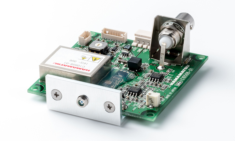
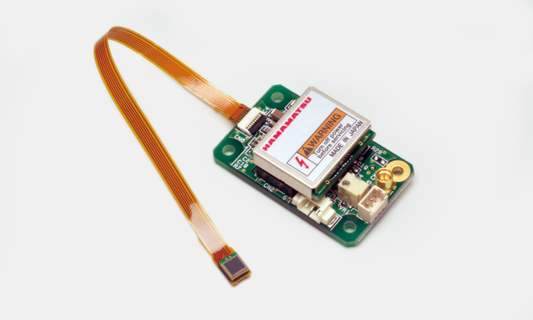
Choosing the right photon detector for each channel
Once light has been split into wavelength bands, the detector choice becomes makes a difference. The same sample can appear different depending on whether a channel is optimized for dim fluorescence sensitivity, for linearity, or for dense multiplexing, so it helps to match PMTs, APDs, and SiPMs to the application.
Photon detection determines sensitivity, dynamic range, and stability for dim fluorescence.
PMT selection guide
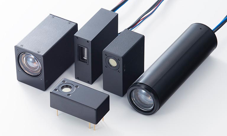
PMTs, APDs, and SiPMs each come with different strengths and trade-offs for flow cytometry, especially when you are balancing dim-fluorescence sensitivity, linearity, and multiplexing.
Where optical performance matters
Flow cytometry generates both physical and biochemical information. Forward scatter (FSC) and side scatter (SSC) report cell size and internal complexity, while fluorescence channels report labelled molecular markers. In multiparametric assays, those readouts are only reliable when excitation is stable, emitted light is cleanly separated, and detector response is matched to the signal level.
This becomes even more important as assays become more complex. Spectral flow cytometry places greater demands on spectral separation, detector channels, and unmixing, while microfluidic cytometry highlights the value of compact optical integration and detector stability in smaller-format systems.
Looking ahead
Flow cytometry’s ability to quantify both physical and molecular characteristics of complex cell populations has made it indispensable across scientific and medical disciplines.
Ongoing technological advances promise further improvements in sensitivity, resolution, and accessibility, ensuring that flow cytometry will remain a cornerstone of modern biological measurement for decades to come.
- Confirmation
-
It looks like you're in the . If this is not your location, please select the correct region or country below.
You're headed to Hamamatsu Photonics website for GB (English). If you want to view an other country's site, the optimized information will be provided by selecting options below.
In order to use this website comfortably, we use cookies. For cookie details please see our cookie policy.
- Cookie Policy
-
This website or its third-party tools use cookies, which are necessary to its functioning and required to achieve the purposes illustrated in this cookie policy. By closing the cookie warning banner, scrolling the page, clicking a link or continuing to browse otherwise, you agree to the use of cookies.
Hamamatsu uses cookies in order to enhance your experience on our website and ensure that our website functions.
You can visit this page at any time to learn more about cookies, get the most up to date information on how we use cookies and manage your cookie settings. We will not use cookies for any purpose other than the ones stated, but please note that we reserve the right to update our cookies.
1. What are cookies?
For modern websites to work according to visitor’s expectations, they need to collect certain basic information about visitors. To do this, a site will create small text files which are placed on visitor’s devices (computer or mobile) - these files are known as cookies when you access a website. Cookies are used in order to make websites function and work efficiently. Cookies are uniquely assigned to each visitor and can only be read by a web server in the domain that issued the cookie to the visitor. Cookies cannot be used to run programs or deliver viruses to a visitor’s device.
Cookies do various jobs which make the visitor’s experience of the internet much smoother and more interactive. For instance, cookies are used to remember the visitor’s preferences on sites they visit often, to remember language preference and to help navigate between pages more efficiently. Much, though not all, of the data collected is anonymous, though some of it is designed to detect browsing patterns and approximate geographical location to improve the visitor experience.
Certain type of cookies may require the data subject’s consent before storing them on the computer.
2. What are the different types of cookies?
This website uses two types of cookies:
- First party cookies. For our website, the first party cookies are controlled and maintained by Hamamatsu. No other parties have access to these cookies.
- Third party cookies. These cookies are implemented by organizations outside Hamamatsu. We do not have access to the data in these cookies, but we use these cookies to improve the overall website experience.
3. How do we use cookies?
This website uses cookies for following purposes:
- Certain cookies are necessary for our website to function. These are strictly necessary cookies and are required to enable website access, support navigation or provide relevant content. These cookies direct you to the correct region or country, and support security and ecommerce. Strictly necessary cookies also enforce your privacy preferences. Without these strictly necessary cookies, much of our website will not function.
- Analytics cookies are used to track website usage. This data enables us to improve our website usability, performance and website administration. In our analytics cookies, we do not store any personal identifying information.
- Functionality cookies. These are used to recognize you when you return to our website. This enables us to personalize our content for you, greet you by name and remember your preferences (for example, your choice of language or region).
- These cookies record your visit to our website, the pages you have visited and the links you have followed. We will use this information to make our website and the advertising displayed on it more relevant to your interests. We may also share this information with third parties for this purpose.
Cookies help us help you. Through the use of cookies, we learn what is important to our visitors and we develop and enhance website content and functionality to support your experience. Much of our website can be accessed if cookies are disabled, however certain website functions may not work. And, we believe your current and future visits will be enhanced if cookies are enabled.
4. Which cookies do we use?
There are two ways to manage cookie preferences.
- You can set your cookie preferences on your device or in your browser.
- You can set your cookie preferences at the website level.
If you don’t want to receive cookies, you can modify your browser so that it notifies you when cookies are sent to it or you can refuse cookies altogether. You can also delete cookies that have already been set.
If you wish to restrict or block web browser cookies which are set on your device then you can do this through your browser settings; the Help function within your browser should tell you how. Alternatively, you may wish to visit www.aboutcookies.org, which contains comprehensive information on how to do this on a wide variety of desktop browsers.
5. What are Internet tags and how do we use them with cookies?
Occasionally, we may use internet tags (also known as action tags, single-pixel GIFs, clear GIFs, invisible GIFs and 1-by-1 GIFs) at this site and may deploy these tags/cookies through a third-party advertising partner or a web analytical service partner which may be located and store the respective information (including your IP-address) in a foreign country. These tags/cookies are placed on both online advertisements that bring users to this site and on different pages of this site. We use this technology to measure the visitors' responses to our sites and the effectiveness of our advertising campaigns (including how many times a page is opened and which information is consulted) as well as to evaluate your use of this website. The third-party partner or the web analytical service partner may be able to collect data about visitors to our and other sites because of these internet tags/cookies, may compose reports regarding the website’s activity for us and may provide further services which are related to the use of the website and the internet. They may provide such information to other parties if there is a legal requirement that they do so, or if they hire the other parties to process information on their behalf.
If you would like more information about web tags and cookies associated with on-line advertising or to opt-out of third-party collection of this information, please visit the Network Advertising Initiative website http://www.networkadvertising.org.
6. Analytics and Advertisement Cookies
We use third-party cookies (such as Google Analytics) to track visitors on our website, to get reports about how visitors use the website and to inform, optimize and serve ads based on someone's past visits to our website.
You may opt-out of Google Analytics cookies by the websites provided by Google:
https://tools.google.com/dlpage/gaoptout?hl=en
As provided in this Privacy Policy (Article 5), you can learn more about opt-out cookies by the website provided by Network Advertising Initiative:
http://www.networkadvertising.org
We inform you that in such case you will not be able to wholly use all functions of our website.
Close