![]()
Products


We are actively taking measures to improve product quality levels.
Applications


Why Hamamatsu?
Resources


Support


Our company


Investors


United States (EN)
Select your region or country.

Light-sheet microscopy
What is light-sheet microscopy?
A light-sheet microscope is an observation technique that illuminates a sample with sheet-shaped light to acquire 2D images. By changing the position where the sheet light is applied, it also enables 3D imaging.
While light-sheet microscopy is often compared to confocal microscopy because it can capture 3D images, it has two significant features:
- Separate optical paths for excitation light illumination and fluorescence detection.
- No excitation light hitting the upper and lower focal planes.
In confocal microscopy, due to the epifluorescence illumination, excitation light hits the upper and lower sides of the focal plane, causing sample fading and phototoxicity. In contrast, light-sheet microscopy illuminates the sample’s focal plane parallelly, avoiding excitation light exposure to the upper and lower sides of the focal plane, which reduces fading and phototoxicity (see Figure 1).
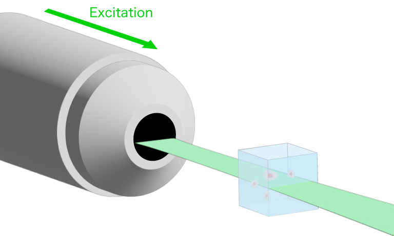
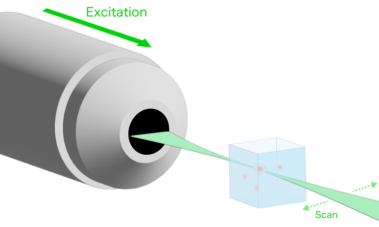
Additionally, while confocal microscopy requires scanning in the XY direction during imaging, light-sheet microscopy illuminates the XY plane all at once, eliminating the need for scanning and enabling faster imaging (see Figures 2 and 3).
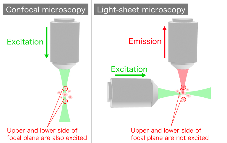
Figure 1: Differences in illumination methods between confocal microscopy and light-sheet microscopy
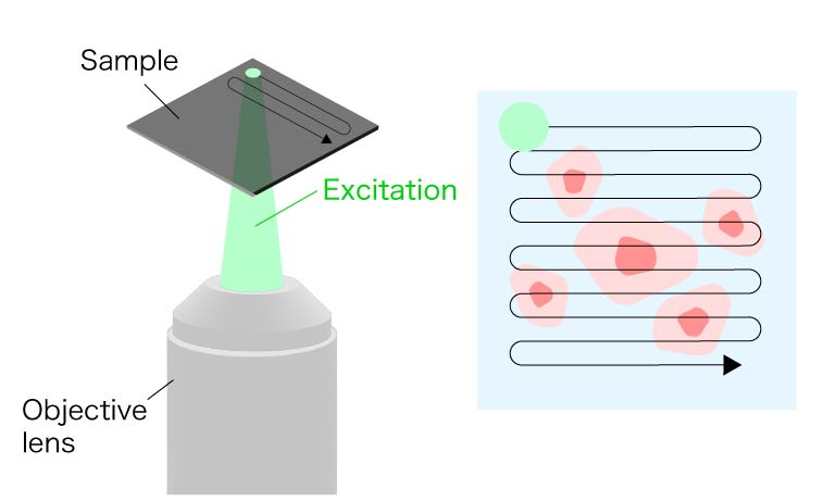
Figure 2: Excitation light illumination image in confocal microscopy
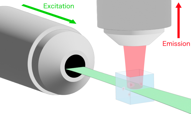
Figure 3: Schematic diagram of light-sheet microscopy (SPIM)
The disadvantage of light-sheet microscopy compared to confocal microscopy lies in its lower resolution. In light-sheet microscopy, you need two objective lenses—one for excitation light illumination and another for fluorescence detection—both of which must be brought close to the sample. However, when attempting to use high-NA (numerical aperture) objective lenses, interference occurs between the objective lenses due to their working distances. As a result, you have no choice but to use low-magnification, low-NA objective lenses with a certain working distance.
Additionally, while sheet light allows clean image acquisition immediately after exiting the excitation objective lens, at positions farther from the emission side of the objective lens, the image quality decreases due to scattering of excitation light by the sample and increased background light. To overcome this, recent advancements include light-sheet microscopes with objective lenses positioned in two directions, enabling excitation light to be irradiated from both directions (see Figure 3).
| Observation method | Confocal microscopy | Light-sheet microscopy |
|---|---|---|
| Capture speed | Slow | Fast |
| Resolution | High | Low |
| Sample phototoxicity | High | Low |
Types of light-sheet microscopy
SPIM(selective plane illumination microscopy)
SPIM (selective plane illumination microscopy) is a light-sheet microscopy technique that creates a sheet of light by expanding a laser using a cylindrical lens. Unlike DSLM (digital scanned light-sheet microscopy), SPIM eliminates the need for laser scanning, resulting in a simpler optical system and easier synchronization with cameras.
DSLM(digital scanned light-sheet microscopy)
DSLM (digital scanned light-sheet microscopy) creates a pseudo-light-sheet by scanning lasers. By combining laser scanning timing with the light-sheet readout mode (mentioned later), DSLM can acquire lower-noise images compared to SPIM.
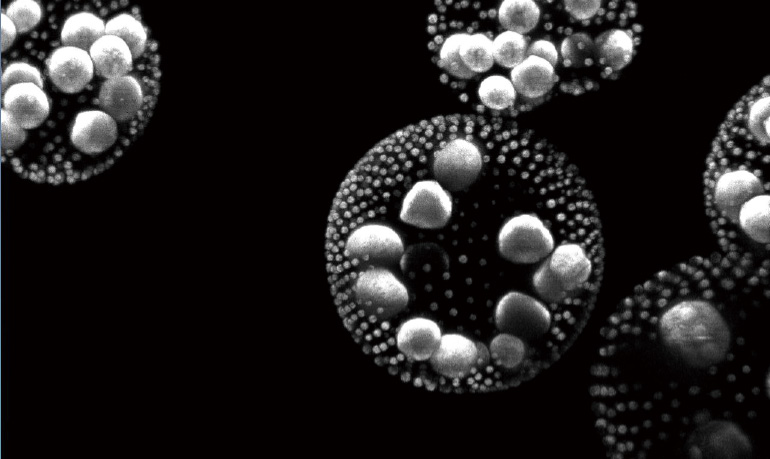
Example images
Free-swimming Volvox carteri scaned by ezDSLM
Data courtesy of Laboratory for Spatiotemporal Regulations Exploratory Research Center on Life and Living Systems (ExCELLS) / National Institute for Basic Biology (NIBB), Dr. Atsushi Taniguchi, Dr. Shigenori Nonaka
Free-swimming Paramecium bursaria scaned by ezDSLM
Data courtesy of Laboratory for Spatiotemporal Regulations Exploratory Research Center on Life and Living Systems (ExCELLS) / National Institute for Basic Biology (NIBB), Dr. Atsushi Taniguchi, Dr. Shigenori Nonaka
GFP-expressing blood cells of Oryzias latipes scanned by ezDSLM
Data courtesy of Laboratory for Spatiotemporal Regulations Exploratory Research Center on Life and Living Systems (ExCELLS) / National Institute for Basic Biology (NIBB), Dr. Atsushi Taniguchi, Dr. Shigenori Nonaka
Sample provided by Spectrography and Bioimaging Facility Core Research Facilities / National Institute for Basic Biology (NIBB), Dr. Joe Sakamoto
3D multicolor observation of Amoeboid movement and the cytoplasm flow
Data courtesy of Laboratory for Spatiotemporal Regulations Exploratory Research Center on Life and Living Systems (ExCELLS) / National Institute for Basic Biology (NIBB), Dr. Atsushi Taniguchi, Dr. Shigenori Nonaka
Quantitative high-speed imaging of developmental processes
Data courtesy of Dr. Philipp L. Keller, Howard Hughes Medical Institute, Janelia Farm Research Campus, Ashburn, Va 20147, USA
Simultaneous two-color imaging of the beating zebrafish heart
Data courtesy of the group of Dr. Jan Huisken from the Max Planck Institute of Molecular Cell Biology and Genetics
Technical introduction: Light-sheet Readout Mode
The Light-sheet Readout Mode is a method for improving the signal-to-noise ratio (S/N) of sCMOS cameras used in light-sheet microscopy. In this mode, it is possible to adjust the timing of camera readout synchronously with the movement of excitation light, allowing for high-quality image acquisition with an unaffected S/N due to scattering.
Hamamatsu Photonics has obtained a patent for this.
Patent numbers:
JP06475307, JP05770958, JP06240056, JP05639670
Recommended products
The ultimate camera, evolved
With 5x faster photon number resolving capabilities, the ORCA-Quest 2 remains the apex camera for ultra low-light, quantitative imaging.
Highlights:
- Ideal for ultra low light and replacing EM-CCDS
- Exclusive quantitative CMOS for pioneering imaging
- Ultra-low noise enables single photon resolution
- High sensitivity does not require speed compromise
This ORCA is ideal for:
- TIRF
- Computational/super-resolution microscopy
- Genetically encoded voltage imaging
- Luminescence
- Quantum computing
- UV applications

Small pixels, big benefit
The ORCA-Fire is a unique back-thinned sCMOS that is optimized for fast, low-mag, large field of view, low-light quantitative imaging.
Highlights:
- Ideal for low light with high pixel volume and speed
- Nyquist sampling at 40x and below
- High sensitivity even at fast speeds
- Advanced bidirectional lightsheet readout modes
This ORCA is ideal for:
- Lightsheet
- Simultaneous multi-wavelength imaging
- High-throughput widefield fluorescence
- Genetically encoded voltage imaging
- Tissue mapping
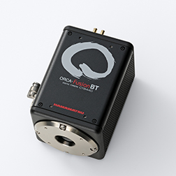
Uncompromising performance
The ORCA-FusionBT sCMOS camera is the perfect synthesis of sensitivity, speed, resoution and overall quantiative, low-light performance.
Highlights:
- Ideal for very low light applications at 60x and 100x
- Superior SNR from maximized QE, minimized noise
- Three speed/noise modes for use-specific imaging
- Exclusive, high QE, back-thinned sCMOS sensor
This ORCA is ideal for:
- Spinning disk confocal
- Lightsheet
- Optogenetics
- Structured illumination microscopy
- Single-molecule localization microscopy
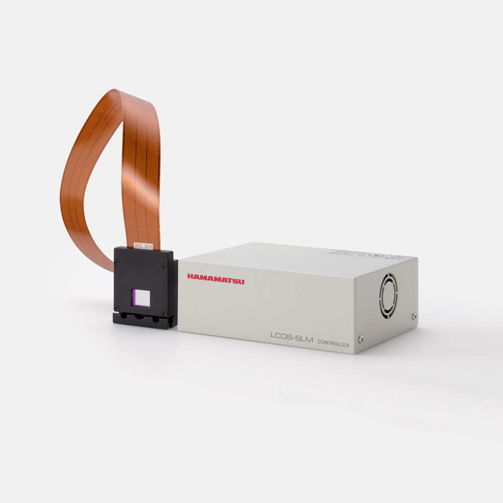
The LCOS-SLM optical phase modulator significantly enhances light-sheet microscopy by enabling precise control over the phase and intensity of the light sheet. This leads to sharper, high-contrast images and minimizes phototoxicity by optimizing light distribution to only target the region of interest.
With its rapid wavefront modulation capabilities, the LCOS-SLM improves image acquisition speed, essential for capturing dynamic biological processes in live samples. Overall, the LCOS-SLM supports research applications with enhanced image quality and reduced sample stress, pushing the state-of-the-art of clarity and efficiency in light-sheet microscopy applications.
- Confirmation
-
It looks like you're in the . If this is not your location, please select the correct region or country below.
You're headed to Hamamatsu Photonics website for US (English). If you want to view an other country's site, the optimized information will be provided by selecting options below.
In order to use this website comfortably, we use cookies. For cookie details please see our cookie policy.
- Cookie Policy
-
This website or its third-party tools use cookies, which are necessary to its functioning and required to achieve the purposes illustrated in this cookie policy. By closing the cookie warning banner, scrolling the page, clicking a link or continuing to browse otherwise, you agree to the use of cookies.
Hamamatsu uses cookies in order to enhance your experience on our website and ensure that our website functions.
You can visit this page at any time to learn more about cookies, get the most up to date information on how we use cookies and manage your cookie settings. We will not use cookies for any purpose other than the ones stated, but please note that we reserve the right to update our cookies.
1. What are cookies?
For modern websites to work according to visitor’s expectations, they need to collect certain basic information about visitors. To do this, a site will create small text files which are placed on visitor’s devices (computer or mobile) - these files are known as cookies when you access a website. Cookies are used in order to make websites function and work efficiently. Cookies are uniquely assigned to each visitor and can only be read by a web server in the domain that issued the cookie to the visitor. Cookies cannot be used to run programs or deliver viruses to a visitor’s device.
Cookies do various jobs which make the visitor’s experience of the internet much smoother and more interactive. For instance, cookies are used to remember the visitor’s preferences on sites they visit often, to remember language preference and to help navigate between pages more efficiently. Much, though not all, of the data collected is anonymous, though some of it is designed to detect browsing patterns and approximate geographical location to improve the visitor experience.
Certain type of cookies may require the data subject’s consent before storing them on the computer.
2. What are the different types of cookies?
This website uses two types of cookies:
- First party cookies. For our website, the first party cookies are controlled and maintained by Hamamatsu. No other parties have access to these cookies.
- Third party cookies. These cookies are implemented by organizations outside Hamamatsu. We do not have access to the data in these cookies, but we use these cookies to improve the overall website experience.
3. How do we use cookies?
This website uses cookies for following purposes:
- Certain cookies are necessary for our website to function. These are strictly necessary cookies and are required to enable website access, support navigation or provide relevant content. These cookies direct you to the correct region or country, and support security and ecommerce. Strictly necessary cookies also enforce your privacy preferences. Without these strictly necessary cookies, much of our website will not function.
- Analytics cookies are used to track website usage. This data enables us to improve our website usability, performance and website administration. In our analytics cookies, we do not store any personal identifying information.
- Functionality cookies. These are used to recognize you when you return to our website. This enables us to personalize our content for you, greet you by name and remember your preferences (for example, your choice of language or region).
- These cookies record your visit to our website, the pages you have visited and the links you have followed. We will use this information to make our website and the advertising displayed on it more relevant to your interests. We may also share this information with third parties for this purpose.
Cookies help us help you. Through the use of cookies, we learn what is important to our visitors and we develop and enhance website content and functionality to support your experience. Much of our website can be accessed if cookies are disabled, however certain website functions may not work. And, we believe your current and future visits will be enhanced if cookies are enabled.
4. Which cookies do we use?
There are two ways to manage cookie preferences.
- You can set your cookie preferences on your device or in your browser.
- You can set your cookie preferences at the website level.
If you don’t want to receive cookies, you can modify your browser so that it notifies you when cookies are sent to it or you can refuse cookies altogether. You can also delete cookies that have already been set.
If you wish to restrict or block web browser cookies which are set on your device then you can do this through your browser settings; the Help function within your browser should tell you how. Alternatively, you may wish to visit www.aboutcookies.org, which contains comprehensive information on how to do this on a wide variety of desktop browsers.
5. What are Internet tags and how do we use them with cookies?
Occasionally, we may use internet tags (also known as action tags, single-pixel GIFs, clear GIFs, invisible GIFs and 1-by-1 GIFs) at this site and may deploy these tags/cookies through a third-party advertising partner or a web analytical service partner which may be located and store the respective information (including your IP-address) in a foreign country. These tags/cookies are placed on both online advertisements that bring users to this site and on different pages of this site. We use this technology to measure the visitors' responses to our sites and the effectiveness of our advertising campaigns (including how many times a page is opened and which information is consulted) as well as to evaluate your use of this website. The third-party partner or the web analytical service partner may be able to collect data about visitors to our and other sites because of these internet tags/cookies, may compose reports regarding the website’s activity for us and may provide further services which are related to the use of the website and the internet. They may provide such information to other parties if there is a legal requirement that they do so, or if they hire the other parties to process information on their behalf.
If you would like more information about web tags and cookies associated with on-line advertising or to opt-out of third-party collection of this information, please visit the Network Advertising Initiative website http://www.networkadvertising.org.
6. Analytics and Advertisement Cookies
We use third-party cookies (such as Google Analytics) to track visitors on our website, to get reports about how visitors use the website and to inform, optimize and serve ads based on someone's past visits to our website.
You may opt-out of Google Analytics cookies by the websites provided by Google:
https://tools.google.com/dlpage/gaoptout?hl=en
As provided in this Privacy Policy (Article 5), you can learn more about opt-out cookies by the website provided by Network Advertising Initiative:
http://www.networkadvertising.org
We inform you that in such case you will not be able to wholly use all functions of our website.
Close