![]()
Products


We are actively taking measures to improve product quality levels.
Applications


Why Hamamatsu?
Resources


Support


Our company


Investors


United States (EN)
Select your region or country.
Photostimulation
What is photostimulation?
Photostimulation refers to an experimental method where samples such as living organisms or cells are stimulated with light, and the resulting reactions are observed. Various experimental techniques utilize photostimulation, including FRAP (fluorescence recovery after photobleaching), optogenetics, photoconversion, and photoswitching. The following sections describe FRAP and optogenetics.
What is FRAP?
FRAP is the measurement of the time required for the fluorescence brightness of a specific fluorescent molecule in the observation area to recover after photobleaching with strong excitation light. It is a method that can estimate the movement speed of molecules from outside the observation area. The less movement of molecules, the slower the recovery speed of fluorescence, and the more movement of molecules, the faster the recovery speed.
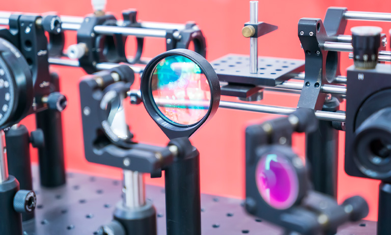
To achieve photobleaching in a specific region, FRAP requires very strong excitation light and optical systems that can precisely target the excitation light to that area. Commonly, lasers used in confocal microscopy are employed for excitation. Spatial light modulators (SLMs) or digital mirror devices (DMDs) are used to focus the laser exclusively on the desired region.
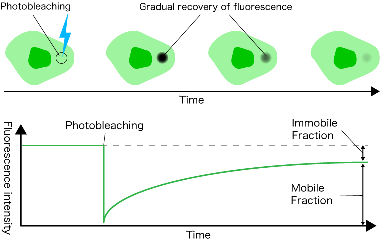
Figure 1: Principle of FRAP
What is optogenetics?
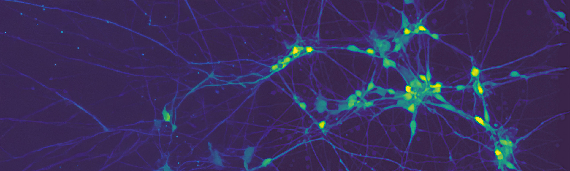
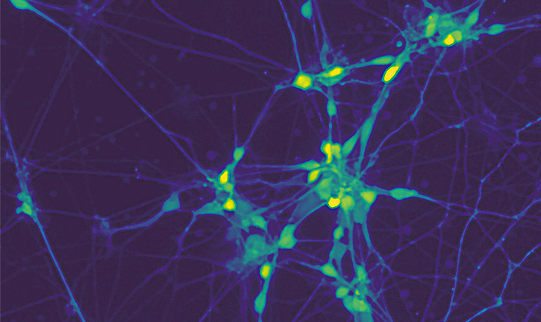
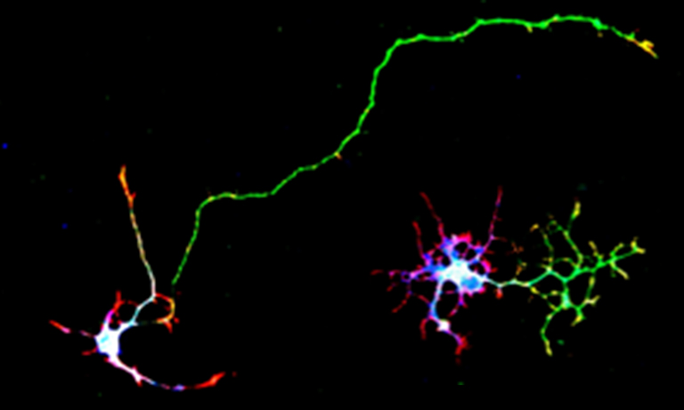
Optogenetics is an interdisciplinary field that combines optics and genetics. It is also known as light genetics. In optogenetics, membrane proteins such as channelrhodopsin and halorhodopsin, which respond to light energy, are expressed in neurons. By irradiating specific wavelengths of light, it is possible to excite or inhibit target neurons. This technique can be applied to elucidate the structure and function of neural networks.
Traditionally, electrical stimulation methods were primarily used to study neural function. However, these methods had a drawback: they stimulated not only the targeted neurons but also neighboring neurons connected to them. Optogenetics overcomes this limitation by allowing precise stimulation of only the neurons expressing membrane proteins like channelrhodopsin. As a result, researchers can observe neural cell movement more accurately.
In optogenetics, there are cases where only specific neurons need to be stimulated with light. In such situations, it is necessary to irradiate light only to specific areas. Additionally, when stimulating multiple neurons simultaneously, light must be directed to multiple areas. Our LCOS-SLM allows customizable illumination patterns, enabling precise light irradiation to specific regions.
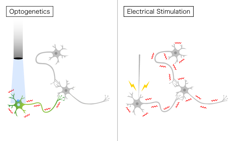
Figure 2: Differences between optogenetics and electrical stimulation
Research introduction: Light-controlled tools for intracellular cAMP generation
In recent years, various light manipulation techniques have made significant advancements in the field of life sciences. Since 2006, light-sensitive ion channels have become an essential tool for optogenetics, the manipulation of light, in the field of neuroscience. However, within living organisms, numerous signals exist beyond ion channels. Notably, cyclic nucleotides such as cAMP and cGMP play crucial roles in diverse intracellular signal transduction pathways. In the main signaling pathway involving cAMP, activation of adenylate cyclase or inhibition of phosphodiesterase leads to an increase in intracellular cAMP concentration, subsequently activating protein kinase A (PKA). This activation is followed by the appearance of voltage-dependent Ca2+ channels and various cellular responses. Hamamatsu Photonics is researching an optogenetics tool called ‘PAC’ that allows light-controlled cAMP generation to induce these responses.
Recommended products
The ultimate camera, evolved
With 5x faster photon number resolving capabilities, the ORCA-Quest 2 remains the apex camera for ultra low-light, quantitative imaging.
Highlights:
- Ideal for ultra low light and replacing EM-CCDS
- Exclusive quantitative CMOS for pioneering imaging
- Ultra-low noise enables single photon resolution
- High sensitivity does not require speed compromise
This ORCA is ideal for:
- TIRF
- Computational/super-resolution microscopy
- Genetically encoded voltage imaging
- Luminescence
- Quantum computing
- UV applications
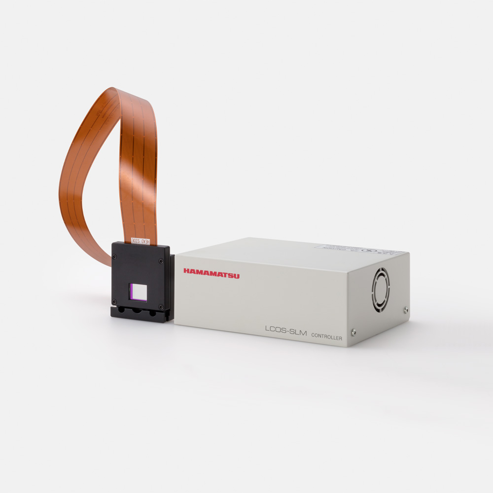
The LCOS-SLM (liquid crystal on silicon - spatial light modulator) is a device that allows electrical control of the phase of laser light. It consists of a structure where a liquid crystal is sandwiched between a CMOS chip with pixel electrodes arranged in a two-dimensional pattern and transparent electrodes deposited on a glass substrate. Digital images output from a PC are converted to analog signals by a dedicated driving circuit and applied with voltage to the pixel electrodes on the CMOS chip.
The LCOS-SLM enables precise controls over light patterns, which enables real-time holographic beam steering for cells, tissues, and microorganisms. Additionally, the LCOS-SLM can create 3D holograms, which are helpfup for target illumination in 3D space for neuroscience, life science, and microscopy applications.
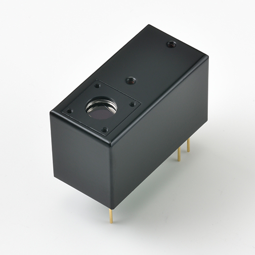
The Hamamatsu H12056-40 is a photomultiplier tube (PMT) module equipped with a built-in gate function, making it ideal for photo stimulation experiments.
In such experiments, there's a risk of detector saturation or damage due to high incident light levels. The H12056-40's gate function allows users to turn off the PMT during periods of intense light exposure, protecting the detector from potential damage and preventing the detection of unwanted light. This feature is particularly beneficial in applications like fluorescence recovery after photobleaching (FRAP) and optogenetics, where precise control over light exposure and detection is crucial.
Additionally, the H12056-40 offers high sensitivity throughout the visible spectrum, enhancing the detection of weak fluorescence signals that follow photostimulation. This combination of high sensitivity and gating capability makes the H12056-40 a great choice for photostimulation setups, ensuring accurate and reliable measurements.
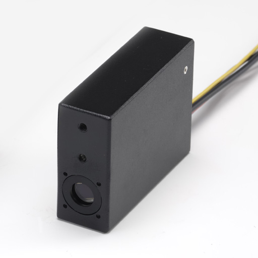
The Hamamatsu H110706-40 is a photomultiplier tube (PMT) module equipped with a built-in gate function, making it ideal for experiments with risk of high light levels, like photostimulation. The H110706-40 's gate function allows users to turn off the PMT, protecting the detector from potential damage and preventing the detection of unwanted light. This feature is particularly beneficial in applications like fluorescence recovery after photobleaching (FRAP) and optogenetics, where precise control over light exposure and detection is crucial.
Additionally, the H110706-40 offers high sensitivity throughout the visible spectrum, enhancing the detection of weak fluorescence signals. This combination of high sensitivity and gating capability makes the H12056-40 a great choice for photostimulation setups, ensuring accurate and reliable measurements.
Our photomultiplier tube (PMT) modules are ideal for photostimulation experiments by providing high sensitivity and low noise, essential for detecting weak fluorescence signals. With fast response times, they support precise, high-speed imaging required for capturing dynamic biological responses. Additionally, Hamamatsu's PMTs feature a broad spectral range, allowing detection of many wavelengths commonly used in photo stimulation setups. This wide spectral range combined with high sensitivity, ensures Hamamatsu’s PMTs deliver the clear, detailed data collection, necessary for accurate, reproducible photostimulation results.
Our Multi-Pixel Photon Counter (MPPC) modules offering high photon detection efficiency and excellent signal-to-noise ratios for photo stimulation experiments. The solid-state design of MPPCs ensures exceptional stability and longevity, with low dark counts and high gain for reliable, repeatable results. In high background environments, they offer higher QE leading to more accurate data. Their compact, durable construction facilitates seamless integration into photostimulation setups. This makes Hamamatsu's MPPC modules an ideal choice for achieving accurate, high-resolution data in photostimulation experiments.
- Confirmation
-
It looks like you're in the . If this is not your location, please select the correct region or country below.
You're headed to Hamamatsu Photonics website for US (English). If you want to view an other country's site, the optimized information will be provided by selecting options below.
In order to use this website comfortably, we use cookies. For cookie details please see our cookie policy.
- Cookie Policy
-
This website or its third-party tools use cookies, which are necessary to its functioning and required to achieve the purposes illustrated in this cookie policy. By closing the cookie warning banner, scrolling the page, clicking a link or continuing to browse otherwise, you agree to the use of cookies.
Hamamatsu uses cookies in order to enhance your experience on our website and ensure that our website functions.
You can visit this page at any time to learn more about cookies, get the most up to date information on how we use cookies and manage your cookie settings. We will not use cookies for any purpose other than the ones stated, but please note that we reserve the right to update our cookies.
1. What are cookies?
For modern websites to work according to visitor’s expectations, they need to collect certain basic information about visitors. To do this, a site will create small text files which are placed on visitor’s devices (computer or mobile) - these files are known as cookies when you access a website. Cookies are used in order to make websites function and work efficiently. Cookies are uniquely assigned to each visitor and can only be read by a web server in the domain that issued the cookie to the visitor. Cookies cannot be used to run programs or deliver viruses to a visitor’s device.
Cookies do various jobs which make the visitor’s experience of the internet much smoother and more interactive. For instance, cookies are used to remember the visitor’s preferences on sites they visit often, to remember language preference and to help navigate between pages more efficiently. Much, though not all, of the data collected is anonymous, though some of it is designed to detect browsing patterns and approximate geographical location to improve the visitor experience.
Certain type of cookies may require the data subject’s consent before storing them on the computer.
2. What are the different types of cookies?
This website uses two types of cookies:
- First party cookies. For our website, the first party cookies are controlled and maintained by Hamamatsu. No other parties have access to these cookies.
- Third party cookies. These cookies are implemented by organizations outside Hamamatsu. We do not have access to the data in these cookies, but we use these cookies to improve the overall website experience.
3. How do we use cookies?
This website uses cookies for following purposes:
- Certain cookies are necessary for our website to function. These are strictly necessary cookies and are required to enable website access, support navigation or provide relevant content. These cookies direct you to the correct region or country, and support security and ecommerce. Strictly necessary cookies also enforce your privacy preferences. Without these strictly necessary cookies, much of our website will not function.
- Analytics cookies are used to track website usage. This data enables us to improve our website usability, performance and website administration. In our analytics cookies, we do not store any personal identifying information.
- Functionality cookies. These are used to recognize you when you return to our website. This enables us to personalize our content for you, greet you by name and remember your preferences (for example, your choice of language or region).
- These cookies record your visit to our website, the pages you have visited and the links you have followed. We will use this information to make our website and the advertising displayed on it more relevant to your interests. We may also share this information with third parties for this purpose.
Cookies help us help you. Through the use of cookies, we learn what is important to our visitors and we develop and enhance website content and functionality to support your experience. Much of our website can be accessed if cookies are disabled, however certain website functions may not work. And, we believe your current and future visits will be enhanced if cookies are enabled.
4. Which cookies do we use?
There are two ways to manage cookie preferences.
- You can set your cookie preferences on your device or in your browser.
- You can set your cookie preferences at the website level.
If you don’t want to receive cookies, you can modify your browser so that it notifies you when cookies are sent to it or you can refuse cookies altogether. You can also delete cookies that have already been set.
If you wish to restrict or block web browser cookies which are set on your device then you can do this through your browser settings; the Help function within your browser should tell you how. Alternatively, you may wish to visit www.aboutcookies.org, which contains comprehensive information on how to do this on a wide variety of desktop browsers.
5. What are Internet tags and how do we use them with cookies?
Occasionally, we may use internet tags (also known as action tags, single-pixel GIFs, clear GIFs, invisible GIFs and 1-by-1 GIFs) at this site and may deploy these tags/cookies through a third-party advertising partner or a web analytical service partner which may be located and store the respective information (including your IP-address) in a foreign country. These tags/cookies are placed on both online advertisements that bring users to this site and on different pages of this site. We use this technology to measure the visitors' responses to our sites and the effectiveness of our advertising campaigns (including how many times a page is opened and which information is consulted) as well as to evaluate your use of this website. The third-party partner or the web analytical service partner may be able to collect data about visitors to our and other sites because of these internet tags/cookies, may compose reports regarding the website’s activity for us and may provide further services which are related to the use of the website and the internet. They may provide such information to other parties if there is a legal requirement that they do so, or if they hire the other parties to process information on their behalf.
If you would like more information about web tags and cookies associated with on-line advertising or to opt-out of third-party collection of this information, please visit the Network Advertising Initiative website http://www.networkadvertising.org.
6. Analytics and Advertisement Cookies
We use third-party cookies (such as Google Analytics) to track visitors on our website, to get reports about how visitors use the website and to inform, optimize and serve ads based on someone's past visits to our website.
You may opt-out of Google Analytics cookies by the websites provided by Google:
https://tools.google.com/dlpage/gaoptout?hl=en
As provided in this Privacy Policy (Article 5), you can learn more about opt-out cookies by the website provided by Network Advertising Initiative:
http://www.networkadvertising.org
We inform you that in such case you will not be able to wholly use all functions of our website.
Close