![]()
Products


We are actively taking measures to improve product quality levels.
Applications


Why Hamamatsu?
Resources


Support


Our company


Investors


United States (EN)
Select your region or country.

Super-resolution microscopy
What is super-resolution microscopy?
Super-resolution microscopy uses a type of microscope that allows observation of samples beyond the diffraction limit typically associated with conventional optical microscopes. The spatial resolution of an ordinary optical microscope is generally calculated using Rayleigh’s resolution formula:
δ= 0.61 × λ / NA
According to this formula, in the visible light range (400 nm to 700 nm), the resolution limit is approximately 162 nm to 284 nm.*1
To surpass this resolution limit, there are several types of super-resolution microscopy have been developed, and the following are explanations of the mechanisms behind different types.
*1: Calculated with an NA (numerical aperture) of 1.5 for the objective lens.
SIM: Structured illumination microscopy
Structured illumination microscopy is a method that obtains super-resolution images by observing Moiré patterns generated when a patterned excitation light called “structured illumination” is applied to the sample and then reconstructing those Moiré images.
Moiré refers to patterns that occur when overlapping periodic motifs. When the sample is illuminated with structured light containing periodic stripe patterns, Moiré interference arises between these patterns and the sample’s fine structures (see Figure 1). Since Moiré images contain information about the fine structures, capturing multiple images with varying structured illumination angles and phases allows computational reconstruction of fine structures, resulting in images with approximately twice the resolution of the original image.
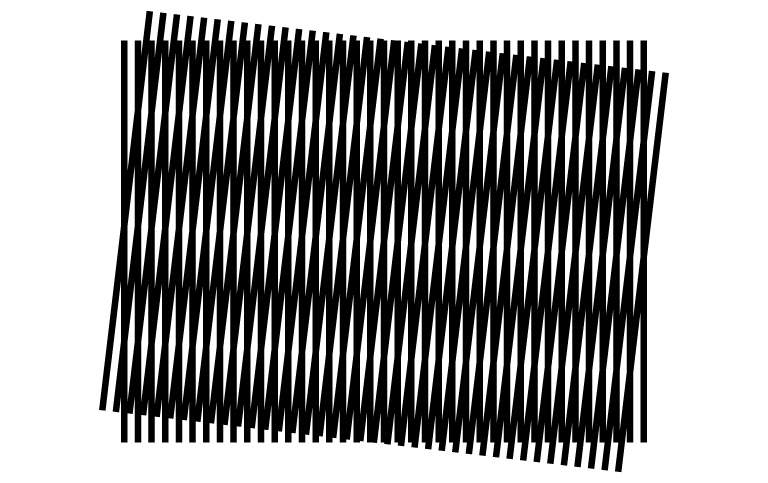
Figure 1: Example of Moiré pattern
PALM/STORM: Localization method
The localization method is a method for generating super-resolution images. It involves randomly illuminating fluorescent molecules and acquiring their centroid positions at the single-molecule level. By overlaying and reconstructing these centroid positions, super-resolution images are obtained.
In conventional optical microscopes, when fluorescent molecules are closer together than the microscope’s resolution limit, their emitted light appears overlapped due to diffraction, making it impossible to distinguish them. However, if we can observe closely spaced molecules individually, we can determine their coordinates within the sample by calculating the centroid positions of their emission points.
In the localization method, the sample is labeled with special fluorescent molecules capable of switching between ON and OFF states. These molecules are randomly activated to emit fluorescence while being repeatedly captured by a camera. The centroid positions of the emission points are then determined from the acquired images, and by performing computational operations on these images, a single super-resolution image is generated (see Figure 2).
There are two approaches within the localization method: PALM (photoactivated localization microscopy) and STORM (stochastic optical reconstruction microscopy). PALM uses fluorescent proteins as labels, while STORM employs synthetic dyes.
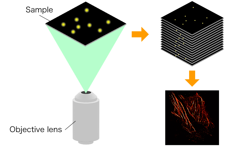
Figure 2: Process of super-resolution image generation using localization method
STED: Stimulated emission depletion
STED (stimulated emission depletion) is a method to obtain super-resolution images by combining two lasers: one for exciting fluorescent molecules and another for deactivating them.
When using lasers to excite fluorescent molecules, the spot size of the laser is determined by its wavelength. If you want to observe fluorescent molecules smaller than the laser spot size, it is necessary to reduce this spot size. However, since the laser spot size is determined by the wavelength, it cannot be physically made smaller (see Figure 3, top).
In STED microscopy, a doughnut-shaped STED light is applied to the fluorescent molecules excited by the excitation laser. This selectively de-excites the excitation light in the peripheral region of the laser spot, allowing only the small central area to emit fluorescence (see Figure 3, bottom). As a result, it becomes possible to detect only the fluorescence within the laser spot size, improving resolution.
While STED microscopy can enhance the XY resolution in the two-dimensional plane using the doughnut-shaped STED light, it does not improve resolution in the Z direction.
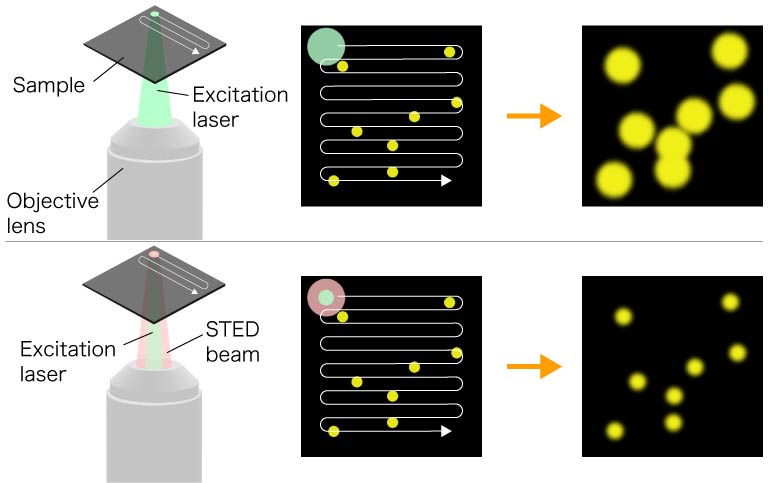
Figure 3: Differences between conventional laser microscopy and STED (stimulated emission depletion) method
Comparison of the methods
| Method | Advantages | Disadvantages |
|---|---|---|
| SIM |
|
|
| PALM/STORM |
|
|
| STED |
|
|
Example images
Super-resolution images using SIM
The ORCAⓇ-Quest 2 qCMOSⓇ camera, with its small pixel size, allows us to obtain higher-resolution images compared to the ORCA-Fusion Digital CMOS camera.
Super-resolution image obtained with ORCA-Quest qCMOS camera
qCMOS camera / 4.6 μm pixel size / Super-resolution system: VT-iSIM
Super-resolution image obtained with ORCA-Fusion Digital CMOS camera
Gen Ⅲ sCMOS camera / 6.5 μm pixel size / Super-resolution system: VT-iSIM
Data courtesy of Steven Coleman (Visitech international Ltd.)
Super-resolution images using localization method
Time lapse super-resolution image of HeLa cells (localization method)
Time lapse STORM image of African green monkey kidney cells (BSC-1)
Recommended products
Regardless of technique, to image below the limit of optical resolution requires precision in the optical system and the use of computational methods to extract the super-resolved image. When errors enter the optical system and/or noise dominates the signal in the image detection, these methods underperform, given less certainty in the quality of the super resolution data. For this reason, the ORCA-Quest2, our flagship quantitative CMOS camera, is the camera of choice. The ultra-quiet noise performance of the ORCA-Quest2 is not just a number on a data sheet. In these experiments this performance reduces errors in the raw image data, leading to more confidence, better information and prettier pictures in the computational results.
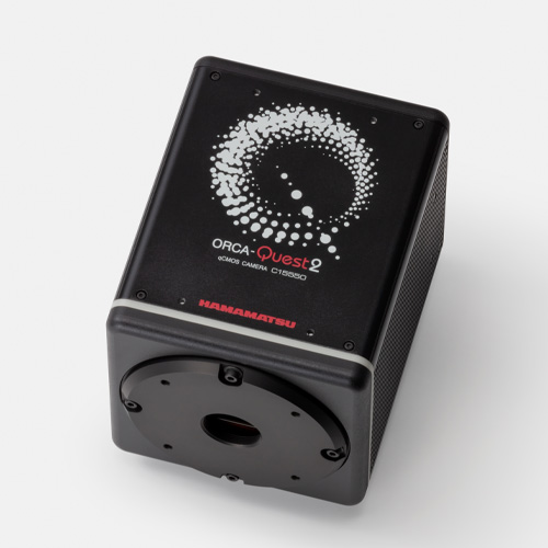
The ultimate camera, evolved
With 5x faster photon number resolving capabilities, the ORCA-Quest 2 remains the apex camera for ultra low-light, quantitative imaging.
Highlights:
- Ideal for ultra low light and replacing EM-CCDS
- Exclusive quantitative CMOS for pioneering imaging
- Ultra-low noise enables single photon resolution
- High sensitivity does not require speed compromise
This ORCA is ideal for:
- TIRF
- Computational/super-resolution microscopy
- Genetically encoded voltage imaging
- Luminescence
- Quantum computing
- UV applications

Small pixels, big benefit
The ORCA-Fire is a unique back-thinned sCMOS that is optimized for fast, low-mag, large field of view, low-light quantitative imaging.
Highlights:
- Ideal for low light with high pixel volume and speed
- Nyquist sampling at 40x and below
- High sensitivity even at fast speeds
- Advanced bidirectional lightsheet readout modes
This ORCA is ideal for:
- Lightsheet
- Simultaneous multi-wavelength imaging
- High-throughput widefield fluorescence
- Genetically encoded voltage imaging
- Tissue mapping
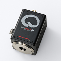
Uncompromising performance
The ORCA-FusionBT sCMOS camera is the perfect synthesis of sensitivity, speed, resoution and overall quantiative, low-light performance.
Highlights:
- Ideal for very low light applications at 60x and 100x
- Superior SNR from maximized QE, minimized noise
- Three speed/noise modes for use-specific imaging
- Exclusive, high QE, back-thinned sCMOS sensor
This ORCA is ideal for:
- Spinning disk confocal
- Lightsheet
- Optogenetics
- Structured illumination microscopy
- Single-molecule localization microscopy
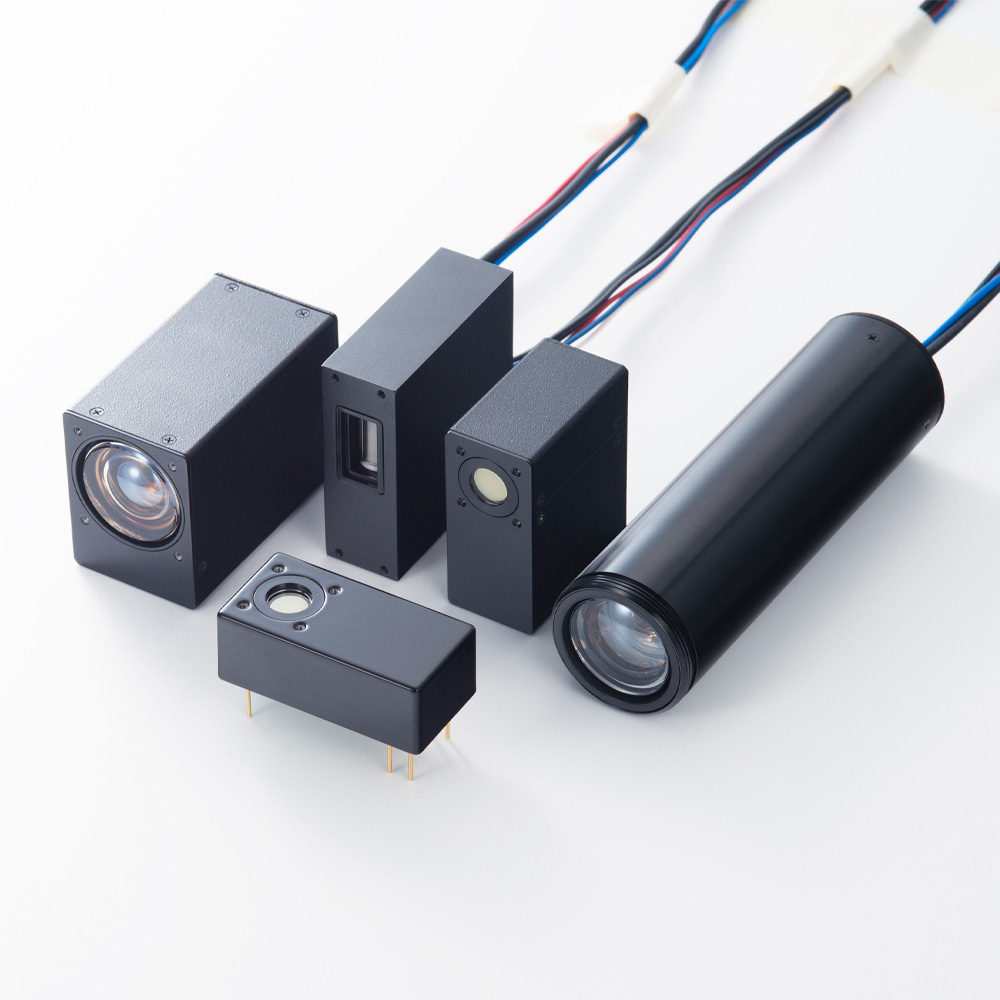
Our photomultiplier tubes (PMTs) empower super-resolution microscopy by delivering unparalleled sensitivity, wide dynamic range and low-noise performance, leading to higher image contrast. Their fast time response allows researchers to image faster, enabling precise analysis of real time processes. By delivering higher sensitivity and faster imaging speeds, Hamamatsu’s PMTs enhance the contrast and speed of data measurements, making them ideal for advanced biomedical research and pushing the boundaries of what's achievable in super-resolution microscopy.
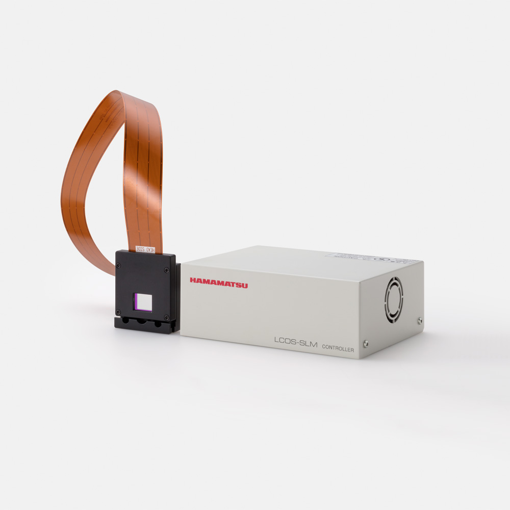
The LCOS-SLM (Liquid Crystal on Silicon - Spatial Light Modulator) is a device that allows electrical control of the phase of laser light. It consists of a structure where a liquid crystal is sandwiched between a CMOS chip with pixel electrodes arranged in a two-dimensional pattern and transparent electrodes deposited on a glass substrate. Digital images output from a PC are converted to analog signals by a dedicated driving circuit and applied with voltage to the pixel electrodes on the CMOS chip.
The LCOS-SLM takes standard laser light, and shapes it into precise patterns of excitation light, crucial for reconstructing high-resolution images, and handle multiple wavelengths for multicolor imaging. The LCOS-SLM optimizes contrast and image clarity, essential for revealing intricate details at the nanoscale. With exceptional phase stability and flexibility, Hamamatsu's LCOS-SLM empowers researchers to push the limits of spatial resolution, facilitating discoveries in cell biology and molecular structures that drive innovation in life sciences and medical research.
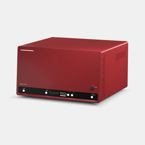
Supercontinuum white light lasers, such as the SuperK series, are ideal for super-resolution microscopy due to their broadband tunability, high spectral power density (SPD), pulsed nature, and single-mode output. These lasers provide bright, diffraction-limited light across a wide spectrum, enabling precise and efficient localization of fluorescent molecules. The pulsed and diffraction-limited nature of the SuperK lasers allows for high temporal and spatial resolution, making them perfect for advanced microscopy techniques.
- Confirmation
-
It looks like you're in the . If this is not your location, please select the correct region or country below.
You're headed to Hamamatsu Photonics website for US (English). If you want to view an other country's site, the optimized information will be provided by selecting options below.
In order to use this website comfortably, we use cookies. For cookie details please see our cookie policy.
- Cookie Policy
-
This website or its third-party tools use cookies, which are necessary to its functioning and required to achieve the purposes illustrated in this cookie policy. By closing the cookie warning banner, scrolling the page, clicking a link or continuing to browse otherwise, you agree to the use of cookies.
Hamamatsu uses cookies in order to enhance your experience on our website and ensure that our website functions.
You can visit this page at any time to learn more about cookies, get the most up to date information on how we use cookies and manage your cookie settings. We will not use cookies for any purpose other than the ones stated, but please note that we reserve the right to update our cookies.
1. What are cookies?
For modern websites to work according to visitor’s expectations, they need to collect certain basic information about visitors. To do this, a site will create small text files which are placed on visitor’s devices (computer or mobile) - these files are known as cookies when you access a website. Cookies are used in order to make websites function and work efficiently. Cookies are uniquely assigned to each visitor and can only be read by a web server in the domain that issued the cookie to the visitor. Cookies cannot be used to run programs or deliver viruses to a visitor’s device.
Cookies do various jobs which make the visitor’s experience of the internet much smoother and more interactive. For instance, cookies are used to remember the visitor’s preferences on sites they visit often, to remember language preference and to help navigate between pages more efficiently. Much, though not all, of the data collected is anonymous, though some of it is designed to detect browsing patterns and approximate geographical location to improve the visitor experience.
Certain type of cookies may require the data subject’s consent before storing them on the computer.
2. What are the different types of cookies?
This website uses two types of cookies:
- First party cookies. For our website, the first party cookies are controlled and maintained by Hamamatsu. No other parties have access to these cookies.
- Third party cookies. These cookies are implemented by organizations outside Hamamatsu. We do not have access to the data in these cookies, but we use these cookies to improve the overall website experience.
3. How do we use cookies?
This website uses cookies for following purposes:
- Certain cookies are necessary for our website to function. These are strictly necessary cookies and are required to enable website access, support navigation or provide relevant content. These cookies direct you to the correct region or country, and support security and ecommerce. Strictly necessary cookies also enforce your privacy preferences. Without these strictly necessary cookies, much of our website will not function.
- Analytics cookies are used to track website usage. This data enables us to improve our website usability, performance and website administration. In our analytics cookies, we do not store any personal identifying information.
- Functionality cookies. These are used to recognize you when you return to our website. This enables us to personalize our content for you, greet you by name and remember your preferences (for example, your choice of language or region).
- These cookies record your visit to our website, the pages you have visited and the links you have followed. We will use this information to make our website and the advertising displayed on it more relevant to your interests. We may also share this information with third parties for this purpose.
Cookies help us help you. Through the use of cookies, we learn what is important to our visitors and we develop and enhance website content and functionality to support your experience. Much of our website can be accessed if cookies are disabled, however certain website functions may not work. And, we believe your current and future visits will be enhanced if cookies are enabled.
4. Which cookies do we use?
There are two ways to manage cookie preferences.
- You can set your cookie preferences on your device or in your browser.
- You can set your cookie preferences at the website level.
If you don’t want to receive cookies, you can modify your browser so that it notifies you when cookies are sent to it or you can refuse cookies altogether. You can also delete cookies that have already been set.
If you wish to restrict or block web browser cookies which are set on your device then you can do this through your browser settings; the Help function within your browser should tell you how. Alternatively, you may wish to visit www.aboutcookies.org, which contains comprehensive information on how to do this on a wide variety of desktop browsers.
5. What are Internet tags and how do we use them with cookies?
Occasionally, we may use internet tags (also known as action tags, single-pixel GIFs, clear GIFs, invisible GIFs and 1-by-1 GIFs) at this site and may deploy these tags/cookies through a third-party advertising partner or a web analytical service partner which may be located and store the respective information (including your IP-address) in a foreign country. These tags/cookies are placed on both online advertisements that bring users to this site and on different pages of this site. We use this technology to measure the visitors' responses to our sites and the effectiveness of our advertising campaigns (including how many times a page is opened and which information is consulted) as well as to evaluate your use of this website. The third-party partner or the web analytical service partner may be able to collect data about visitors to our and other sites because of these internet tags/cookies, may compose reports regarding the website’s activity for us and may provide further services which are related to the use of the website and the internet. They may provide such information to other parties if there is a legal requirement that they do so, or if they hire the other parties to process information on their behalf.
If you would like more information about web tags and cookies associated with on-line advertising or to opt-out of third-party collection of this information, please visit the Network Advertising Initiative website http://www.networkadvertising.org.
6. Analytics and Advertisement Cookies
We use third-party cookies (such as Google Analytics) to track visitors on our website, to get reports about how visitors use the website and to inform, optimize and serve ads based on someone's past visits to our website.
You may opt-out of Google Analytics cookies by the websites provided by Google:
https://tools.google.com/dlpage/gaoptout?hl=en
As provided in this Privacy Policy (Article 5), you can learn more about opt-out cookies by the website provided by Network Advertising Initiative:
http://www.networkadvertising.org
We inform you that in such case you will not be able to wholly use all functions of our website.
Close