![]()
Products


We are actively taking measures to improve product quality levels.
Applications


Why Hamamatsu?
Resources


Support


Our company


Investors


United States (EN)
Select your region or country.
Multiphoton microscopy
What is multiphoton microscopy?
Multiphoton microscopy is a type of laser scanning fluorescence microscope represented by confocal microscopy. It utilizes long-wavelength, ultrashort pulse lasers and the phenomenon of multiphoton excitation to observe the deep regions of a sample.
The main advantages of multiphoton microscopy are as follows:
- Excitation light is of a long wavelength, allowing observation of deep regions.
- Only fluorescent dye molecules near the focal point are excited, minimizing fading.
- Fluorescence occurs only at the focal plane, eliminating the need for a pinhole and efficiently detecting scattered or refracted fluorescence within the sample.
- Due to its blueshift property, it enables simultaneous observation of multiple wavelengths.
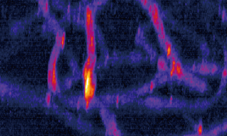
Two-photon microscope image of mouse brain nerve cell
Provided by: Department of Medical Spectroscopy (now BioPhotonics Innovation Laboratory), Hamamatsu University School of Medicine
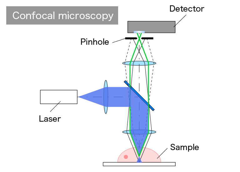
Figure 1: Optical system schematic of a confocal microscope
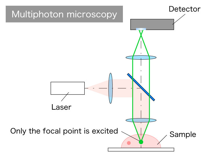
Figure 2: Optical system schematic of a multiphoton microscope
What is multiphoton excitation?
Multiphoton excitation refers to the phenomenon where fluorescent molecules are excited by simultaneously absorbing multiple photons. In this section, two-photon excitation, in which two photons are absorbed simultaneously, is used as an example.
Normally, if a fluorescent molecule is excited by 500 nm light, it absorbs a single photon at that wavelength and enters an excited state. However, in the case of two-photon excitation, it absorbs two photons with a wavelength of 1000 nm (which is twice the 500 nm wavelength) and becomes excited. Since the energy intensity of a single photon is inversely proportional to its wavelength, if we consider the energy of a 1000 nm photon as 1, the energy of a 500 nm photon would be 2. By absorbing two 1000 nm photons, the fluorescent molecule reaches the same excitation energy level.
To induce multiphoton excitation, extremely high photon density (intensity) is required momentarily. Therefore, femtosecond-level ultrashort pulse lasers are commonly used. Multiphoton excitation is a rare phenomenon, typically occurring only in the vicinity of the focal point where photons are concentrated by the objective lens.
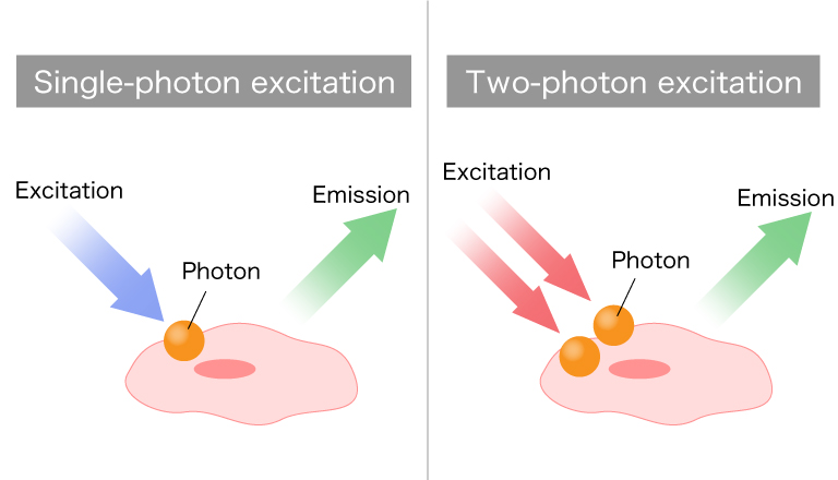
Figure 3: Concept of two-photon excitation
What is blueshift?
Blueshift is the property where the maximum absorption wavelength of excitation light, for example during two-photon excitation, shifts to shorter wavelengths compared to the ideal wavelength.
For instance, when performing two-photon excitation with 1000 nm excitation light, it would be ideal for the excitation to occur at 500 nm. However, in practice, blueshift can cause excitation at wavelengths below 500 nm. This property allows simultaneous excitation of multiple fluorescent dyes using a single excitation wavelength.
While conventional fluorescence microscopes require multiple light sources or excitation filters to excite different fluorescent molecules individually, the application of blueshift in multiphoton microscopy enables observation of multiple wavelengths simultaneously using a single laser.
Example images
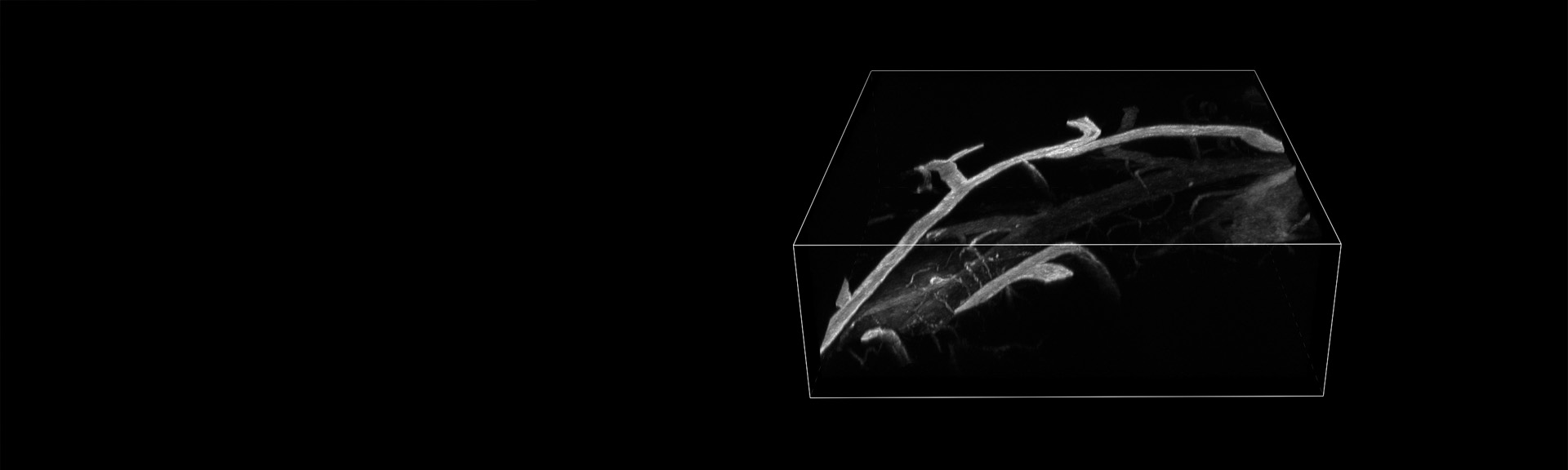
Observation of the mouse brain’s deep regions
Using a Hamamatsu Photonics photomultiplier tube module equivalent to H15460-40
Provided by: Masanori Murayama, Ph.D., RIKEN Center for Brain Science
Observation of glomeruli in pathological mouse models (HIGA)
Three-dimensional fluorescence observation of kidneys removed from each of the HIGA mouse model of IGA nephropathy (left) and wild-type mice (right) using two-photon microscopy. Blue: vascular endothelium, Green: basement membrane, Red: podocyte secondary process. In the HIGA mouse model of IGA nephropathy, the basement membrane between the vascular endothelium and the podocyte secondary process is seen to bulge out like a spherical ball. In wild-type mice, on the other hand, the basement membrane is not bulged.
Provided by: Aru Konno, Associate Professor, Department of Microbiology and Immunology, Hamamatsu University School of Medicine
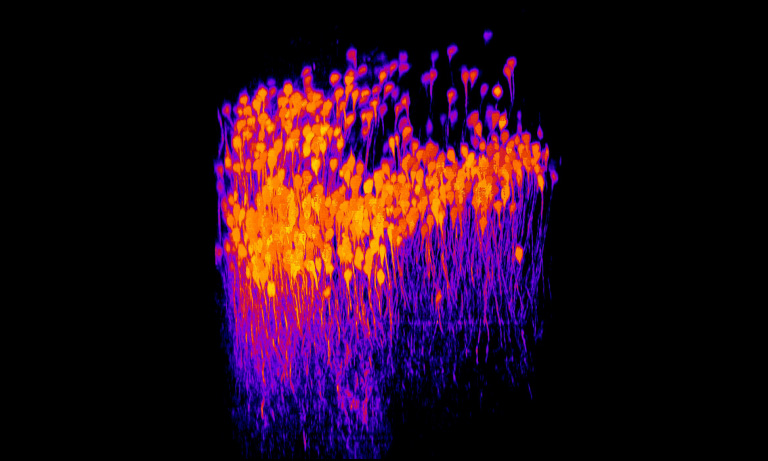
High resolution observation of Thy1-YFP mouse using SLM two-photon microscopy
A cross-section of a brain sample from a Thy1-YFP mouse was observed with a NA1.1 objective lens for two-photon observation. By correcting astigmatism and coma aberration that exist in the optical system with an SLM, cell bodies can be observed with high resolution.
Provided by: Aru Konno, Associate Professor, Department of Microbiology and Immunology, Hamamatsu University School of Medicine
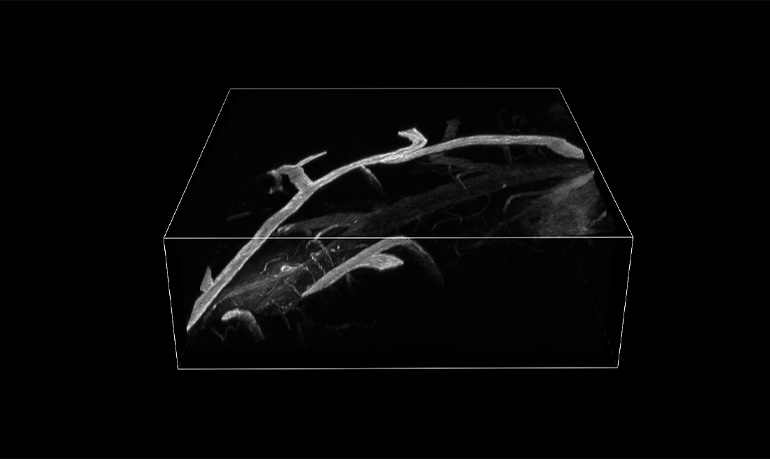
3D image of brain nerve fibers in Thy1-YFP mice using SLM two-photon microscopy
This image is a 3D reconstruction of a cross-section of a brain sample from a Thy1-YFP mouse, observed with a two-photon NA1.1 objective lens. By correcting astigmatism and coma aberration of the optical system with an SLM, cell bodies can be observed at high resolution and projections of brain nerve fibers can be clearly observed.
Provided by: Aru Konno, Associate Professor, Department of Microbiology and Immunology, Hamamatsu University School of Medicine
Multipoint scanning with SLM
HeLa cells were observed using a laser-scanning two-photon microscope equipped with an SLM. The left image shows four simultaneous excitation points using the SLM, whereas the right image shows a conventional single-point scan. 4-point simultaneous excitation enables 4-fold faster imaging. The SLM also enables us to obtain images comparable to single-point scanning even with four-point scanning by aligning the intensities among multiple points.
Aberration correction by SLM
3 µm beads encapsulated in epoxy resin were observed using a water immersion objective lens with NA1.0. 200 µm x 200 µm in the XY direction was scanned using a galvanometer scanner, and 1000 images were acquired in the Z direction by moving the objective lens by 1.5 µm. Without SLM correction, spherical aberration occurs due to the difference in refractive index between water and epoxy resin. This causes the focused light shape to elongate vertically at deeper depths, reducing the energy density and making it difficult to observe the beads at deeper depths. On the other hand, SLM correction improves the focusing shape of the excitation light and maintains a high energy density even in deep areas.
Research introduction: High-performance multiphoton excitation microscopy using SLM
By using a spatial light modulator (SLM) to precisely control the wavefront of light, it is possible to manipulate light to improve the performance and functions of an optical system, such as forming multiple focal points to enable simultaneous multipoint observation and correcting optical distortion (aberration), which is a cause of reduced resolution. We aim to incorporate this SLM into a multiphoton excitation fluorescence microscope system to control the wavefront of the excitation laser light to enable highly accurate and simple observation of a living body from the surface to its depths. Currently, we are conducting research and development focused on achieving high precision and functionality using a two-photon excitation fluorescence microscope. We also collaborate with Hamamatsu University School of Medicine to explore fundamental and applied research using this advanced microscope system. Our goal is to contribute to medical and biological research by partnering with various universities in the future.
Recommended products
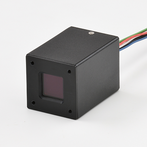
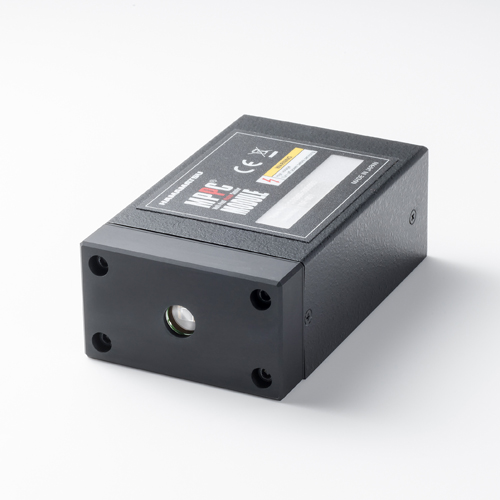
The H15460-40 is a photomultiplier tube module that employs a GaAsP photocathode photomultiplier tube. It has a wide effective surface area of 14 mm × 14 mm, high gain, and high sensitivity. This makes it an optimal detector for applications such as multiphoton excitation microscopy, where extremely weak light needs to be detected.
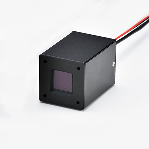
Multiphoton microscopy is a technique that allows accurate imaging of deep structures. However, as the depth increases, the fluorescence captured by small detectors is reduced due. The H15460-40 has a significantly larger active area compared to conventional photomultiplier tubes, enabling the capture of the lowest levels of fluorescence that cannot be captured by typical photomultiplier tubes. The H15461-40 contributions to high signal-to-noise ratio (S/N) image acquisition in multiphoton microscopy.
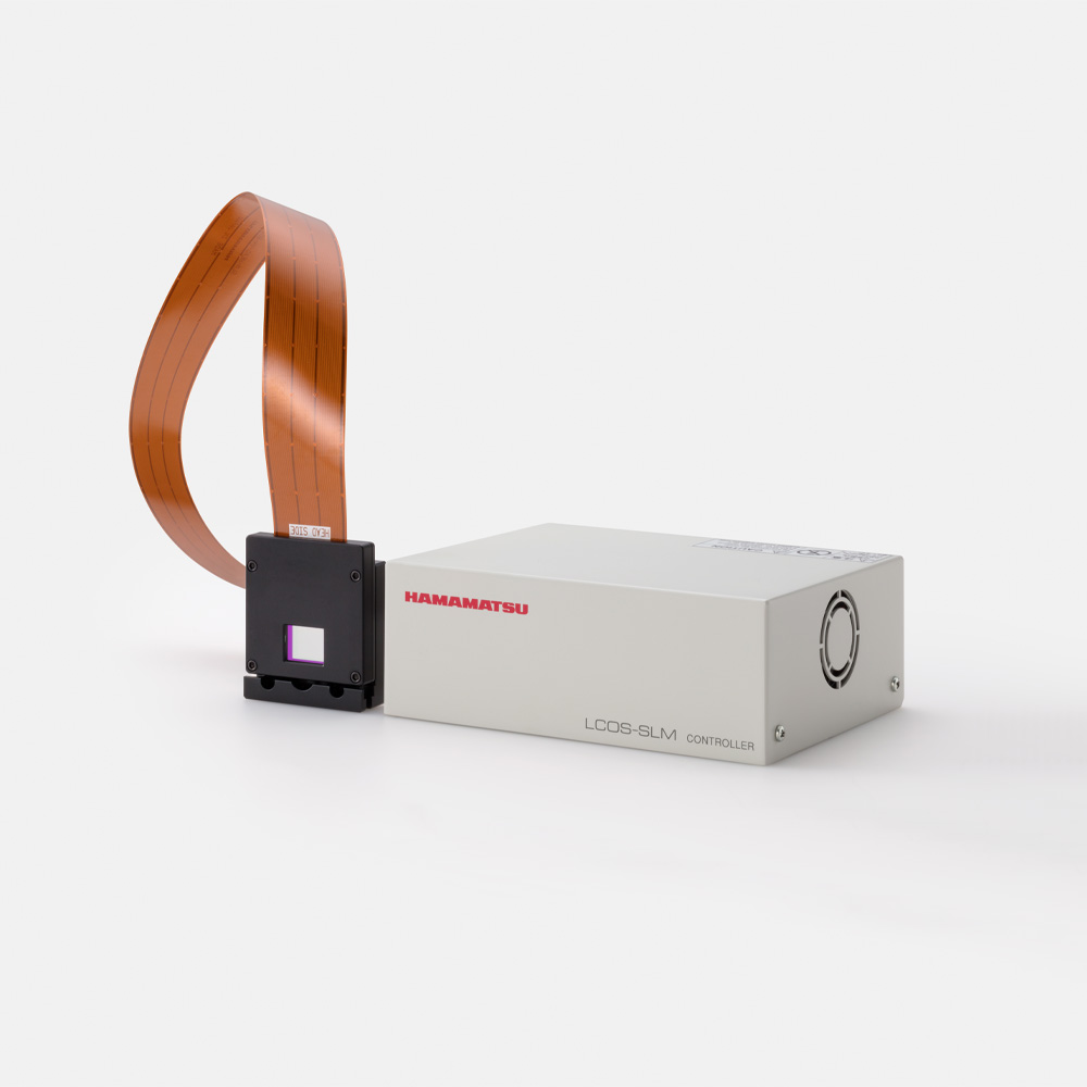
The LCOS-SLM (Liquid Crystal on Silicon - Spatial Light Modulator) is a device that electrically controls the phase of laser light by using a liquid crystal layer between a CMOS chip and transparent electrodes, enabling precise light phase manipulation through voltage application. It is utilized in various applications, including aberration correction, multipoint scanning, and resolution improvement in multiphoton microscopy.
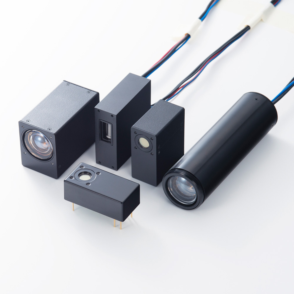
Our photomultiplier tube (PMT) modules modules enhance multiphoton microscopy by delivering exceptional sensitivity and low noise, leading to higher contrast when detecting the faint fluorescence signals. Their fast response time allows faster imaging speeds, capturing dynamic biological processes with precision. Additionally, our PMTs offer a wide spectral response range enabling detection of multiple fluorophores in one sample. This adaptability, combined with reliable performance in demanding research settings, makes Hamamatsu's PMTs invaluable for achieving higher contrast while improving imaging speed in multiphoton microscopy applications.
Our Multi-Pixel Photon Counter (MPPC) modules are ideal for multiphoton microscopy by delivering high photon detection efficiency, and excellent signal-to-noise ratios, ideal for low-light imaging applications. With their solid-state design, MPPCs offer exceptional stability and longevity, providing reliable performance with low dark counts, no damage from high light levels, and high gain. Their compact, robust structure allows easy integration into microscope setups, enhancing sensitivity for capturing fine details, and fast biological processes in deep tissue imaging. Additionally, the gain mechanisms of the MPPC leads to a low excess noise factor allowing photon number resolution to be achieved. This makes Hamamatsu’s MPPC modules a powerful choice for achieving high-resolution, precise results in multiphoton microscopy applications.
- Confirmation
-
It looks like you're in the . If this is not your location, please select the correct region or country below.
You're headed to Hamamatsu Photonics website for US (English). If you want to view an other country's site, the optimized information will be provided by selecting options below.
In order to use this website comfortably, we use cookies. For cookie details please see our cookie policy.
- Cookie Policy
-
This website or its third-party tools use cookies, which are necessary to its functioning and required to achieve the purposes illustrated in this cookie policy. By closing the cookie warning banner, scrolling the page, clicking a link or continuing to browse otherwise, you agree to the use of cookies.
Hamamatsu uses cookies in order to enhance your experience on our website and ensure that our website functions.
You can visit this page at any time to learn more about cookies, get the most up to date information on how we use cookies and manage your cookie settings. We will not use cookies for any purpose other than the ones stated, but please note that we reserve the right to update our cookies.
1. What are cookies?
For modern websites to work according to visitor’s expectations, they need to collect certain basic information about visitors. To do this, a site will create small text files which are placed on visitor’s devices (computer or mobile) - these files are known as cookies when you access a website. Cookies are used in order to make websites function and work efficiently. Cookies are uniquely assigned to each visitor and can only be read by a web server in the domain that issued the cookie to the visitor. Cookies cannot be used to run programs or deliver viruses to a visitor’s device.
Cookies do various jobs which make the visitor’s experience of the internet much smoother and more interactive. For instance, cookies are used to remember the visitor’s preferences on sites they visit often, to remember language preference and to help navigate between pages more efficiently. Much, though not all, of the data collected is anonymous, though some of it is designed to detect browsing patterns and approximate geographical location to improve the visitor experience.
Certain type of cookies may require the data subject’s consent before storing them on the computer.
2. What are the different types of cookies?
This website uses two types of cookies:
- First party cookies. For our website, the first party cookies are controlled and maintained by Hamamatsu. No other parties have access to these cookies.
- Third party cookies. These cookies are implemented by organizations outside Hamamatsu. We do not have access to the data in these cookies, but we use these cookies to improve the overall website experience.
3. How do we use cookies?
This website uses cookies for following purposes:
- Certain cookies are necessary for our website to function. These are strictly necessary cookies and are required to enable website access, support navigation or provide relevant content. These cookies direct you to the correct region or country, and support security and ecommerce. Strictly necessary cookies also enforce your privacy preferences. Without these strictly necessary cookies, much of our website will not function.
- Analytics cookies are used to track website usage. This data enables us to improve our website usability, performance and website administration. In our analytics cookies, we do not store any personal identifying information.
- Functionality cookies. These are used to recognize you when you return to our website. This enables us to personalize our content for you, greet you by name and remember your preferences (for example, your choice of language or region).
- These cookies record your visit to our website, the pages you have visited and the links you have followed. We will use this information to make our website and the advertising displayed on it more relevant to your interests. We may also share this information with third parties for this purpose.
Cookies help us help you. Through the use of cookies, we learn what is important to our visitors and we develop and enhance website content and functionality to support your experience. Much of our website can be accessed if cookies are disabled, however certain website functions may not work. And, we believe your current and future visits will be enhanced if cookies are enabled.
4. Which cookies do we use?
There are two ways to manage cookie preferences.
- You can set your cookie preferences on your device or in your browser.
- You can set your cookie preferences at the website level.
If you don’t want to receive cookies, you can modify your browser so that it notifies you when cookies are sent to it or you can refuse cookies altogether. You can also delete cookies that have already been set.
If you wish to restrict or block web browser cookies which are set on your device then you can do this through your browser settings; the Help function within your browser should tell you how. Alternatively, you may wish to visit www.aboutcookies.org, which contains comprehensive information on how to do this on a wide variety of desktop browsers.
5. What are Internet tags and how do we use them with cookies?
Occasionally, we may use internet tags (also known as action tags, single-pixel GIFs, clear GIFs, invisible GIFs and 1-by-1 GIFs) at this site and may deploy these tags/cookies through a third-party advertising partner or a web analytical service partner which may be located and store the respective information (including your IP-address) in a foreign country. These tags/cookies are placed on both online advertisements that bring users to this site and on different pages of this site. We use this technology to measure the visitors' responses to our sites and the effectiveness of our advertising campaigns (including how many times a page is opened and which information is consulted) as well as to evaluate your use of this website. The third-party partner or the web analytical service partner may be able to collect data about visitors to our and other sites because of these internet tags/cookies, may compose reports regarding the website’s activity for us and may provide further services which are related to the use of the website and the internet. They may provide such information to other parties if there is a legal requirement that they do so, or if they hire the other parties to process information on their behalf.
If you would like more information about web tags and cookies associated with on-line advertising or to opt-out of third-party collection of this information, please visit the Network Advertising Initiative website http://www.networkadvertising.org.
6. Analytics and Advertisement Cookies
We use third-party cookies (such as Google Analytics) to track visitors on our website, to get reports about how visitors use the website and to inform, optimize and serve ads based on someone's past visits to our website.
You may opt-out of Google Analytics cookies by the websites provided by Google:
https://tools.google.com/dlpage/gaoptout?hl=en
As provided in this Privacy Policy (Article 5), you can learn more about opt-out cookies by the website provided by Network Advertising Initiative:
http://www.networkadvertising.org
We inform you that in such case you will not be able to wholly use all functions of our website.
Close