![]()
Products


We are actively taking measures to improve product quality levels.
Applications


Why Hamamatsu?
Resources


Support


Our company


Investors


United States (EN)
Select your region or country.
3D imaging of neurons cultured in microfluidic devices
Published on February 25, 2025
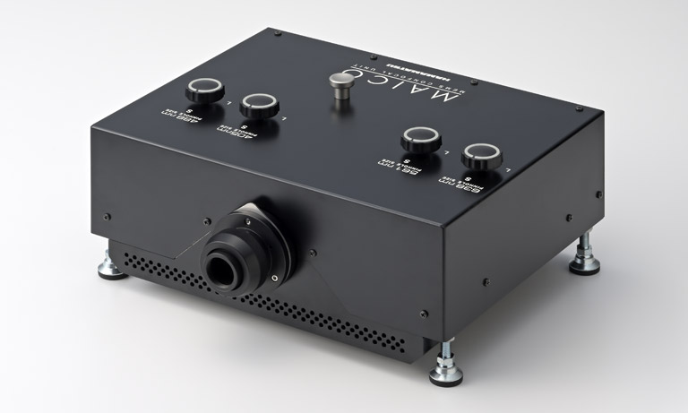
The Nano-Integration Devices and Systems Laboratory at the Research Institute of Electrical Communication (RIEC), Tohoku University, specializes in brain-inspired non-von Neumann computing and the foundational technologies for related hardware research. Within this laboratory, Associate Professor Hideaki Yamamoto's research group, the Nano-Integration Neurocomputing Systems Group, combines semiconductor microfabrication, nerve cell culture, and mathematical modeling to develop new in vitro systems for bottom-up analysis of brain functions. In these in vitro systems, cultured neurons occasionally aggregate to form a 3D structure. The group introduced the MAICO® MEMS confocal unit for 3D imaging of these aggregated neurons.
We interviewed Prof. Hideaki Yamamoto and Mr. Hakuba Murota, who is responsible for cell imaging and analysis using the MAICO MEMS confocal unit, to learn about the background of implementing the MAICO MEMS confocal unit, their experience using it, and future research prospects.
- Current research
- Challenges in Neuronal Imaging
- Decisive factor for introducing the MAICO MEMS confocal unit
- Usability of the MAICO MEMS confocal unit
- Imaging examples
- Prospects for research
- About Multicellular Neurobiocomputing: Understanding and Advancing towards Biological Supremacy
- Researcher profile
Current research
Could you tell us about your research?
Our current research focuses on developing new in vitro systems that can serve as models for the complex network of neurons that constitute animal brains. For example, while research on the heart or cancer has advanced to the point where physiological functions and pathological conditions can be reproduced using cultured cells, there are currently no effective model systems for extremely complex tissues like the brain, where we see a critical issue. For instance, when nerve cells obtained from rat cortices are cultured on a flat dish, they form a network that exhibit activity patterns different from those in living tissue. Therefore, one of our research motivations is to make these activity patterns more like to those in brain tissue. Our group has a background in electronics. Therefore, while new cell culture technologies like organoids are rapidly advancing, we want to realize the goal by using semiconductor manufacturing technologies to reproduce local wiring structures in the brain with living cells.
Specifically, using equipment in our semiconductor clean room (Figure 1), we create microfluidic devices that guide nerve cells to adhere to and extend their neurites (Figure 2). When neural cells are cultured in microfluidic devices, they form dense connections within each cell (through-hole) and then extend their neurites through microchannels connecting the cells to form neural networks. This structure - where densely connected cell populations weakly interact with each other - is said to be a characteristic of actual cerebral cortical neural circuits. By culturing neural cells using this device, we are conducting experiments to partially reproduce network structures similar to those seen in the brain.
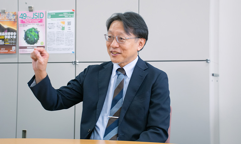
Associate professor Hideki Yamamoto
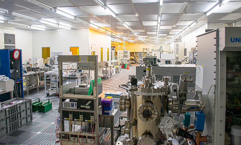
Figure 1: Clean room where microfluidic devices and other devices are fabricated
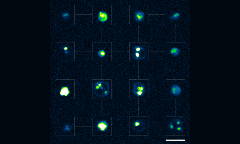
Figure 2: Neurons cultured in a microfluidic device
Challenges in Neuronal Imaging
Mr. Hakuba Murota
What are the challenges of imaging neurons?
When we image cultured nerve cells, we typically use an epifluorescence microscope with a sCMOS camera attached. However, when cell culture continues for several days, region with high cell density form aggregates and develop into 3D structures. With the conventional combination of an epifluorescence microscope and a sCMOS camera, we couldn't image these structures in 3D. As a result, our group had been exploring culture methods to minimize cell aggregation. However, if the system could function well even with aggregated cells, we wanted to keep this characteristic.
This created the need for a confocal microscope to observe these 3D structures, but at the time, our laboratory didn't have a confocal microscope, nor was there one available in our facility's shared equipment. While confocal microscopes were available at other campuses, transporting live samples were cumbersome, so we wanted a confocal microscope that we could use within our research facility. We faced a dilemma, because confocal microscopes are generally expensive, making it difficult to acquire one without securing a substantial budget.
Decisive factor for introducing the MAICO MEMS confocal unit
What made you decide to introduce MAICO?
When we were facing the problem of not having a confocal microscope in our laboratory, we came across information at an exhibition about a new unit from Hamamatsu Photonics that could be attached to an existing microscope to build a confocal microscope by. This caught our interest, and in the following year, we arranged a product demonstration. With its satisfactory sensitivity and image quality, we immediately decided to adopt it.
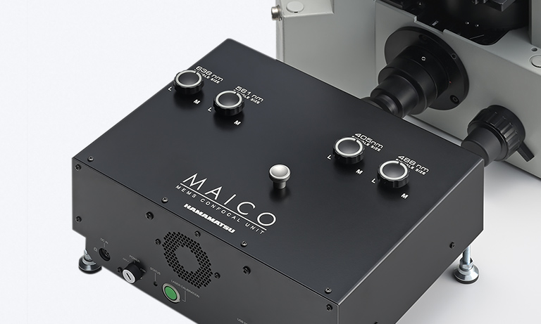
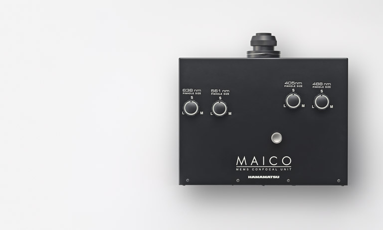
The most decisive factor was the reasonable price. Our laboratory initially introduced the unit with a single 488 nm wavelength configuration. We remember being very grateful for the budget-friendly aspect as it cost less than 5 million yen, which was extremely reasonable. Additionally, we appreciated that it had a subunit structure allowing us to add necessary wavelengths as needed. Currently, we are operating with a two-wavelength configuration after adding a 638 nm unit. Looking ahead, we are considering adding 405 nm and 561 nm units, depending on the progress of our research.
Usability of the MAICO MEMS confocal unit
How would you feel about the usability of MAICO?
We haven't extensively used confocal microscopes from other manufacturers, so we can't make direct comparisons, but we find it attractive that we can start imaging in about 15 minutes after powering on the main unit. The HCImage software used to control MAICO is also intuitive and easy to use. Additionally, the resolution simulator provided by Hamamatsu Photonics is nice, which allows us to easily check in advance the achievable resolutions.
Currently, we are using MAICO for imaging aggregated neurons and are very satisfied with the clear visualization of their 3D structures. Furthermore, even for neurons growing in 2D, MAICO provides higher-resolution compared to a standard camera. While we are currently using a sCMOS camera for calcium imaging of neurons, MAICO could potentially be used for calcium imaging of aggregates in the future, given its fast frame rate.
Imaging examples
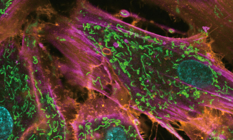
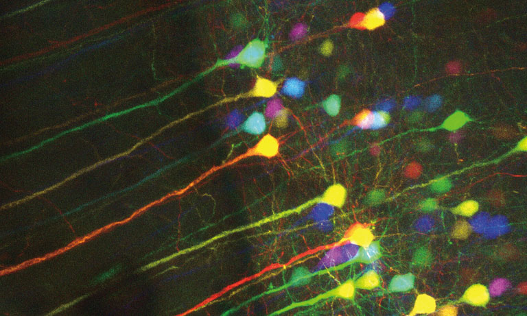
3D images of aggregated rat cortical neurons (imaging NeuO and GCaMP6s fluorescence).
Data provided by: Hideaki Yamamoto, Nano-Integration Devices and Systems Laboratory for Nanoelectronics and Spintronics, Research Institute of Electrical Communication, Tohoku University
Axons and dendrites in cultured rat cortical neurons.
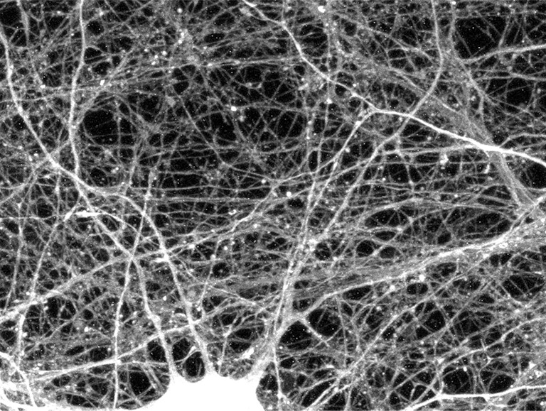
This is a max projection image of neurons captured outside a microfluidic device.
Data provided by: Hideaki Yamamoto, Nano-Integration Devices and Systems Laboratory for Nanoelectronics and Spintronics, Research Institute of Electrical Communication, Tohoku University
Prospects for research
Could you tell us about future research prospects?
The first goal is to increase the number of wells in the microfluidic device to construct a more complex network. Previously, we cultured neurons in a 2 × 2 well array, but now we have created a microfluidic device with a 4 × 4 well array. By increasing the number of wells and thus the number of neurons, we are trying to replicate more complex neuronal networks. Additionally, in the future, we may create 3D microfluidic devices with microchannels arranged not only in the XY direction but also in the Z direction. Although creating 3D structures with the current fabrication process remains challenging, the ability to perform 3D observations with a confocal microscope encourages us to take on such challenge.
The second goal is calcium imaging of neuronal aggregates. Until now, we have primarily performed calcium imaging of neurons cultured 2D, either on cover glasses or within microfluidic devices. However, with access to MAICO, we now have the capability to perform calcium imaging on aggregated neurons. If this works well, it could open new directions in our research, such as actively culturing neurons in aggregates.

About Multicellular Neurobiocomputing: Understanding and Advancing towards Biological Supremacy
Could you tell us about the newly launched “Multicellular Neurobiocomputing: Understanding and Advancing towards Biological Supremacy” project?
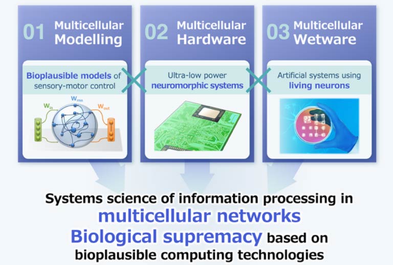
This project was launched in April 2024 with support from the Grant-in-Aid for Transformative Research Areas (A). The phrase "Advancing towards Biological Supremacy" in the project title signifies the idea that systems inspired by biological mechanisms can solve specific problems with learning efficiency, energy efficiency, and environmental adaptability that are difficult to achieve with conventional computers. As I mentioned earlier, our brain is formed by neurons that are connected to create networks. Unlike transistors, or the elements of integrated circuits in computers and smartphones, neurons are inherently unstable. However, our brain performs complex information processing autonomously and with high energy efficiency. This property does not arise from a single cell and cannot be explained simply as a sum of individual elements. Instead, brain functions emerge through the sophisticated arrangement and wiring of various types of neurons, forming a multicellular network. In this project, our goal is to understand how the brain uses networks of biological elements to process infromation by combining mathematical modeling with in vivo and in vitro experiments, and translate this understanding into practical system applications. This research is expected to contribute not only to a fundamental understanding of the nervous system but also to the development of innovative computing technologies with high computational efficiency, robustness, and adaptability.
To achieve this, the project brings together researchers from diverse fields, including information science, bioengineering, biology, and electronics. We have set three key reserch domains: Multicellular Modeling (the formulation of information processing models and learning rules based on biological experiments), Multicellular Hardware (the implementation of functions in hardware and application to robotics), and Multicellular Wetware (the verification of mathematical models and learning rules using cultured cells and the artificial reconstruction of biological functions). Through these domains, we are challenging to demonstrate Biological Supremacy, aiming to harness the principles of biology in next-generation information and communication technology.
Researcher profile
Hideaki Yamamoto
Associate Professor, RIEC, Tohoku University
Associate Professor, Advanced Institute for Materials Research (AIMR), Tohoku University
Associate Professor, Department of Electrical, Information and Physics Engineering, Tohoku University
Associate Professor, Department of Electronic Engineering, Graduate School of Engineering, Tohoku University
Mar. 2009
Ph.D., Department of Nanoscience and Nanoengineering, Graduate School of Advanced Science and Engineering, Waseda University
Apr. 2009
JSPS Research Fellowship for Young Scientists (PD), Waseda University
Apr. 2010
JSPS Research Fellowship for Young Scientists (SPD), Tokyo University of Agriculture and Technology
Apr. 2013
Assistant Professor, Waseda Institute for Advanced Study (WIAS)
Apr. 2014
Assistant Professor, Frontier Research Institute for Interdisciplinary Sciences (FRIS), Tohoku University
May. 2018
Assistant Professor, AIMR, Tohoku University
Jan. 2020
Current Role
Hakuba Murota
Ph.D., Nano-Bio Hybrid Molecular Devices Laboratory, RIEC, Tohoku University
Mar. 2022
Bachelor's Degree in Engineering, Department of Electrical, Information and Physics Engineering, Tohoku University
Mar. 2024
Master's Degree in Engineering, Department of Electronic Engineering, Graduate School of Engineering, Tohoku University
Apr. 2024
Doctoral course, Department of Electronic Engineering, Graduate School of Engineering, Tohoku University
*The content presented on this page is based on an interview conducted in October 2024.
Other case studies
The Laboratory for Evolutionary Cell Biology of Skin, Cosmetics Course, School of Bioscience and Biotechnology, Tokyo University of Technology, is researching epidermal barrier formation mechanisms. To elucidate the mechanisms of epidermal barrier formation, it is necessary to image the epidermis in three dimensions. For this purpose, they have introduced our MAICO® MEMS confocal Unit.
We interviewed Professor. Takeshi Matsui from the same laboratory about the background of introducing the MAICO MEMS confocal unit, his impressions of its use, and the prospects for future research.
MAICO enables imaging with reduced bleed-through between wavelengths, which is an issue in multi-wavelength simultaneous observation. We will introduce how we have achieved a reduction of bleed-through.
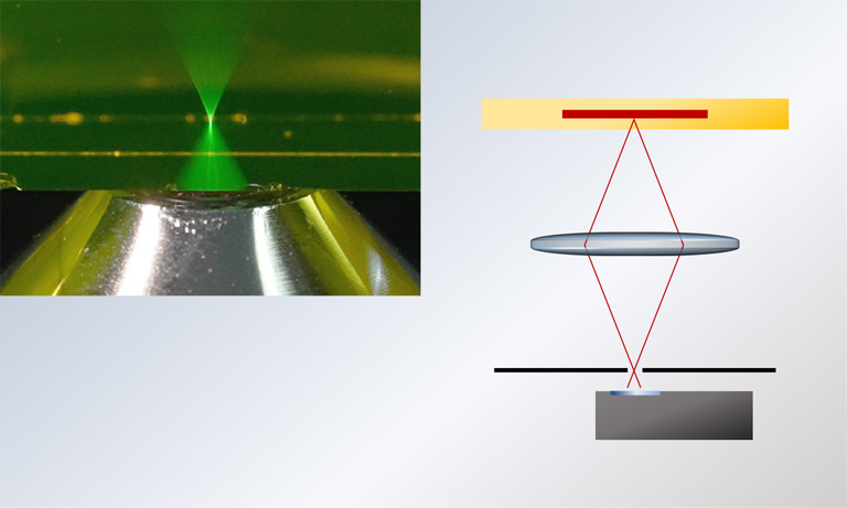
Explanation of the principles of a confocal microscope, which enables you to acquire an image that is less blurry, higher contrast, and higher resolution.

- Confirmation
-
It looks like you're in the . If this is not your location, please select the correct region or country below.
You're headed to Hamamatsu Photonics website for US (English). If you want to view an other country's site, the optimized information will be provided by selecting options below.
In order to use this website comfortably, we use cookies. For cookie details please see our cookie policy.
- Cookie Policy
-
This website or its third-party tools use cookies, which are necessary to its functioning and required to achieve the purposes illustrated in this cookie policy. By closing the cookie warning banner, scrolling the page, clicking a link or continuing to browse otherwise, you agree to the use of cookies.
Hamamatsu uses cookies in order to enhance your experience on our website and ensure that our website functions.
You can visit this page at any time to learn more about cookies, get the most up to date information on how we use cookies and manage your cookie settings. We will not use cookies for any purpose other than the ones stated, but please note that we reserve the right to update our cookies.
1. What are cookies?
For modern websites to work according to visitor’s expectations, they need to collect certain basic information about visitors. To do this, a site will create small text files which are placed on visitor’s devices (computer or mobile) - these files are known as cookies when you access a website. Cookies are used in order to make websites function and work efficiently. Cookies are uniquely assigned to each visitor and can only be read by a web server in the domain that issued the cookie to the visitor. Cookies cannot be used to run programs or deliver viruses to a visitor’s device.
Cookies do various jobs which make the visitor’s experience of the internet much smoother and more interactive. For instance, cookies are used to remember the visitor’s preferences on sites they visit often, to remember language preference and to help navigate between pages more efficiently. Much, though not all, of the data collected is anonymous, though some of it is designed to detect browsing patterns and approximate geographical location to improve the visitor experience.
Certain type of cookies may require the data subject’s consent before storing them on the computer.
2. What are the different types of cookies?
This website uses two types of cookies:
- First party cookies. For our website, the first party cookies are controlled and maintained by Hamamatsu. No other parties have access to these cookies.
- Third party cookies. These cookies are implemented by organizations outside Hamamatsu. We do not have access to the data in these cookies, but we use these cookies to improve the overall website experience.
3. How do we use cookies?
This website uses cookies for following purposes:
- Certain cookies are necessary for our website to function. These are strictly necessary cookies and are required to enable website access, support navigation or provide relevant content. These cookies direct you to the correct region or country, and support security and ecommerce. Strictly necessary cookies also enforce your privacy preferences. Without these strictly necessary cookies, much of our website will not function.
- Analytics cookies are used to track website usage. This data enables us to improve our website usability, performance and website administration. In our analytics cookies, we do not store any personal identifying information.
- Functionality cookies. These are used to recognize you when you return to our website. This enables us to personalize our content for you, greet you by name and remember your preferences (for example, your choice of language or region).
- These cookies record your visit to our website, the pages you have visited and the links you have followed. We will use this information to make our website and the advertising displayed on it more relevant to your interests. We may also share this information with third parties for this purpose.
Cookies help us help you. Through the use of cookies, we learn what is important to our visitors and we develop and enhance website content and functionality to support your experience. Much of our website can be accessed if cookies are disabled, however certain website functions may not work. And, we believe your current and future visits will be enhanced if cookies are enabled.
4. Which cookies do we use?
There are two ways to manage cookie preferences.
- You can set your cookie preferences on your device or in your browser.
- You can set your cookie preferences at the website level.
If you don’t want to receive cookies, you can modify your browser so that it notifies you when cookies are sent to it or you can refuse cookies altogether. You can also delete cookies that have already been set.
If you wish to restrict or block web browser cookies which are set on your device then you can do this through your browser settings; the Help function within your browser should tell you how. Alternatively, you may wish to visit www.aboutcookies.org, which contains comprehensive information on how to do this on a wide variety of desktop browsers.
5. What are Internet tags and how do we use them with cookies?
Occasionally, we may use internet tags (also known as action tags, single-pixel GIFs, clear GIFs, invisible GIFs and 1-by-1 GIFs) at this site and may deploy these tags/cookies through a third-party advertising partner or a web analytical service partner which may be located and store the respective information (including your IP-address) in a foreign country. These tags/cookies are placed on both online advertisements that bring users to this site and on different pages of this site. We use this technology to measure the visitors' responses to our sites and the effectiveness of our advertising campaigns (including how many times a page is opened and which information is consulted) as well as to evaluate your use of this website. The third-party partner or the web analytical service partner may be able to collect data about visitors to our and other sites because of these internet tags/cookies, may compose reports regarding the website’s activity for us and may provide further services which are related to the use of the website and the internet. They may provide such information to other parties if there is a legal requirement that they do so, or if they hire the other parties to process information on their behalf.
If you would like more information about web tags and cookies associated with on-line advertising or to opt-out of third-party collection of this information, please visit the Network Advertising Initiative website http://www.networkadvertising.org.
6. Analytics and Advertisement Cookies
We use third-party cookies (such as Google Analytics) to track visitors on our website, to get reports about how visitors use the website and to inform, optimize and serve ads based on someone's past visits to our website.
You may opt-out of Google Analytics cookies by the websites provided by Google:
https://tools.google.com/dlpage/gaoptout?hl=en
As provided in this Privacy Policy (Article 5), you can learn more about opt-out cookies by the website provided by Network Advertising Initiative:
http://www.networkadvertising.org
We inform you that in such case you will not be able to wholly use all functions of our website.
Close