
Products










We are actively taking measures to improve product quality levels.
Applications












Why Hamamatsu?
Resources





Support




Our company






Investors







United Kingdom (EN)
Select your region or country.

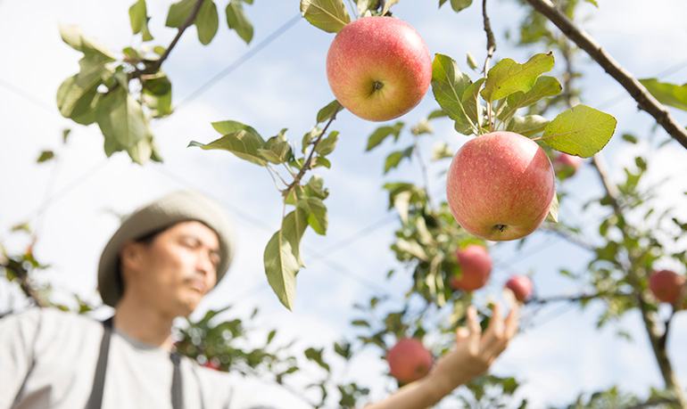
Component Analysis of Crops
Component Analysis of Crops
Component analysis of crops plays an extremely important role in the research and development of crop varieties as well as pesticides and fertilizers. Elucidating the chemical components contained in crops and their physiological functions is expected to promote research and development in areas such as variety improvement, the development of pesticides and fertilizers and the optimization of application conditions, thereby contributing to the improvement of crop productivity and quality. A wide range of analytical methods are used for this purpose, including spectroscopic methods and mass spectrometry. In addition to the development of analytical instruments, sample preparation processes such as extraction and separation have become increasingly sophisticated over the years, enabling the highly sensitive detection and quantitative analysis of trace components.
The Need for Imaging Analysis in Crop Research
Spatial information on chemical components is generally lost during preparation processes such as extraction and separation. Therefore, the importance of imaging analysis for visualizing this information is increasing. In research on plants, including crops, understanding “where” and “in what state” contained components exist serves as a crucial foundation for elucidating physiological functions and mechanisms of action. This is expected to promote research and development in areas such as variety improvement, the development of pesticides and fertilizers, and the optimization of application conditions, thereby contributing to improved crop productivity and quality. In particular, mass spectrometry imaging (MSI), based on mass spectrometry, has attracted attention in recent years as a label-free imaging technique that enables the simultaneous visualization of the spatial distribution of different molecular species while leveraging its high sensitivity.
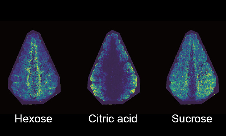
Strawberry component imaging (MSI image)
Comparison of analytical methods for pesticide analysis
The analysis of pesticide-related components is essential for safety assessment of crops and the development of new pesticides. Here, as an example, we compare the characteristics of representative analytical methods and mass spectrometry imaging used for the analysis of pesticide components.
Chromatography-mass spectrometry
(LC/MS, GC/MS, etc.)

Liquid chromatography-mass spectrometry (LC/MS) and gas chromatography-mass spectrometry (GC/MS) are methods that can simultaneously analyze multiple components, such as pesticides and their metabolites, with high sensitivity. First, components are separated using various chromatography methods. Then, the separated compounds are ionized and detected with high sensitivity using instruments such as tandem mass spectrometers, which are well suited for trace analysis. While this method is ideal for monitoring pesticide residues, spatial information cannot be obtained as sample homogenization is required during extraction and purification.
Radioisotope imaging
(RI imaging)
RI imaging is a method that analyzes the migration pathways and distribution of the compound within a plant by administering a substance labeled with a radioactive isotope (RI) to the plant and tracking the radiation emitted by the RI using instruments such as autoradiography, PETIS, and RRIS. This method enables highly sensitive detection, and depending on the method, real-time changes can also be observed. On the other hand, there are practical limitations, such as the inherent inability to distinguish between pesticide active ingredients and their metabolites, the cost of isotope-labeled reagents, and the safety management and maintenance of dedicated facilities resulting from the use of radioactive materials.
Mass spectrometry imaging
(MSI)
Mass spectrometry imaging (MSI) is a technique that acquires mass spectra linked to the spatial information of the sample to be measured, thereby visualizing the distribution of components. It does not require labeling for detection and can simultaneously analyze multiple components, such as pesticides and metabolites. On the other hand, it is important to note that quantitative analysis faces challenges due to variations in sample preparation conditions and the distribution of impurities within the analysis area. Furthermore, MSI measurements require thinly sectioned samples.
| Comparison items | LC/MS, GC/MS | RI imaging | MS imaging |
|---|---|---|---|
| Main applications | Trace and quantitative analysis, qualitative analysis | Tracking of labeled molecules | Distribution analysis of multiple components |
| Bulk analysis/Imaging | Bulk analysis | Imaging | Imaging |
| Labeled/Unlabeled | Unlabeled | Labeled | Unlabeled |
| Destructive/Non-destructive | Destructive | Non-destructive | Destructive |
| Advantages | Established protocols | Live imaging available | Visualization of unlabeled molecules |
| Challenges | Lack of spatial information | Management of RI facilities | Thin sectioning is not suitable for some crop samples |
Challenges of MSI in crop component analysis
When performing MSI, frozen samples must be pre-sectioned to a thickness of several tens of micrometers. For agricultural products with high moisture content or curved leaf surfaces, the sample often collapses during thin sectioning, making the thin sectioning itself extremely difficult. This sample preparation requirement for MSI, which relies on thin sectioning, is considered one of the factors limiting the widespread adoption of MSI in this field.
Hamamatsu Photonics has focused on this challenge and has been developing technologies to realize an alternative sample preparation method to thin sectioning.

What is Poropare™ Transfer plate?
Poropare Transfer plate is a sample preparation support item for MSI, developed by Hamamatsu Photonics’ proprietary technology. As an alternative to thin sectioning, which is a challenge for agricultural samples, it enables an approach called transfer sampling, which transfers components onto the plate. This makes MSI applicable to many crop samples that were previously difficult to analyze because thin sectioning was not feasible.
Poropare’s high water absorbency allows for the transfer of components onto the plate while minimizing analyte delocalization, even for samples with high moisture content. Furthermore, the robust plate enables even hard samples such as leaves to be sampled by pressing onto the plate. Additionally, as the process is completed simply by pressing the sample surface onto the plate, stable sample preparation can be achieved regardless of operator skill.
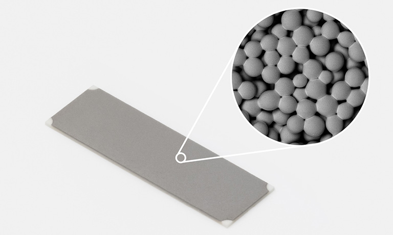
Poropare™ Transfer plate A15551 series
Case study 1: Imaging the components of strawberries

A cross-section of a 50 mm strawberry was pressed onto Poropare to transfer its components without thin sectioning. After the transfer, MSI analysis was performed on the Poropare without any additional processing. Conventional MSI requires the sample to be sectioned to a thickness of tens of micrometers, which not only relies on operator skill but also requires dedicated equipment, creating a significant barrier to implementing MSI. Poropare eliminates the need for such complex sample preparation, enabling transfer sampling that preserves spatial information simply by lightly pressing the cross-section of the sample.
Poropare clearly visualized the distribution of sugars and organic acids within the strawberry. A key advantage of Poropare is that it enables analysis of differences among specific regions such as the peel, flesh, and vascular bundles, which are difficult to capture by conventional bulk analysis that yields only averaged information for the whole fruit, while still allowing the entire fruit to be visualized at a scale close to direct visual observation.
Measurement procedure

1. Expose the measurement surface

2. Press and transfer

3. Obtain measurement results
Case study 2: Evaluation of Pesticide Penetration and Translocation in Leaves
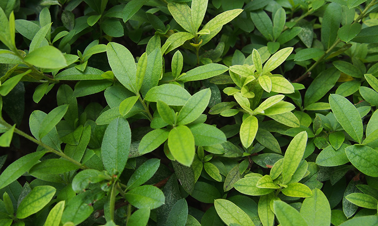
As leaves are thin and curved, they are difficult to section, and it has therefore been considered difficult to apply MSI to obtain component distributions along the leaf surface. Even with approaches where samples are directly introduced into the instrument for measurement, there is concern that the unevenness and curvature of the leaf surface may affect the analysis results and the instrument itself. In this case study, to address these challenges, the entire leaf, on which the pesticide had been applied locally, was crushed on Poropare to perform transfer sampling. After the transfer, MSI analysis was performed on the Poropare without any additional processing.
MSI using Poropare enabled the visualization of pesticide distribution across the entire leaf surface, contributing to the evaluation of pesticide penetration and translocation. It was confirmed that, even under conditions where pressure-induced disruption of the distribution might be a concern, the localized distribution could be preserved during transfer due to the water absorbency of the Poropare. This demonstrates the possibility of performing component imaging of the leaf surface, which was difficult with conventional methods, more simply and stably.
Compared with the conventional RI imaging method, this approach does not require labeling and is therefore free from the constraints associated with RI labeling, reducing the barrier to analysis. In addition, while RI imaging has difficulty distinguishing between pesticide active ingredients and their metabolites due to its principle of detecting label-derived signals, this challenge can be overcome by using label-free MSI, which can simultaneously detect multiple components.
Measurement procedure

1. Apply pesticide and wait for several days

2. Crush the leaf surface
*Image is for illustrative purposes only.

3. Obtain measurement results
Measurement results provided by Nissan Chemical Corporation
The future of crop analysis through mass spectrometry imaging
1. Towards analysis supporting the field of variety improvement and cultivation research
MSI is a method that allows for simultaneous, label-free analysis of various components contained in crops. By reducing the effort required to measure each component individually and capturing a wide range of internal information from crop samples, it offers a new perspective for variety improvement and cultivation research. Transfer sampling using Poropare simplifies sample collection for MSI and reduces reliance on specialized sample preparation procedures. For example, by facilitating sample collection at remote locations or cultivation sites and easing the burden of sample preparation at analytical facilities, it makes MSI more accessible for research and evaluation.


2. A clue to identifying changes within crops
MSI allows us to determine where pesticides, their metabolites, and beneficial components are located within crops. This makes it easier to identify differences that cannot be seen from average component levels alone, and the relationships among multiple components, deepening the understanding of changes occurring within crop samples. Poropare facilitates the handling of samples that were difficult to process with conventional MSI, allowing more practical access to such information across a wide range of crops. As a result, it is expected to support research aimed at increasing the added value of crops, such as understanding the characteristics of each variety and exploring functional components.
3. Supporting better agricultural decision-making
The multi-component imaging information provided by MSI offers new insights for fundamental research on plant functions, analysis of the mechanisms of action and metabolism of pesticides, and the optimization of cultivation conditions. By revealing not only average values across the whole crop, but also the spatial distribution of components, it allows for more specific comparisons and evaluations. Using Poropare makes it easier to handle many diverse samples, facilitating the accumulation of comparable data. As a result, the new data may contribute to reducing the environmental impact of pesticides and enhancing cultivation management. Furthermore, these evaluation data are expected to be utilized as one of the criteria for decision-making in smart agriculture and to create synergistic effects with other technologies.
Guide to purchasing Poropare
Poropare is a product designed for measurements using mass spectrometers equipped with MALDI and DESI ion sources.
Poropare is currently available at the following companies.
*Poropare is a registered trademark of Hamamatsu Photonics K.K.
- Confirmation
-
It looks like you're in the . If this is not your location, please select the correct region or country below.
You're headed to Hamamatsu Photonics website for GB (English). If you want to view an other country's site, the optimized information will be provided by selecting options below.
In order to use this website comfortably, we use cookies. For cookie details please see our cookie policy.
- Cookie Policy
-
This website or its third-party tools use cookies, which are necessary to its functioning and required to achieve the purposes illustrated in this cookie policy. By closing the cookie warning banner, scrolling the page, clicking a link or continuing to browse otherwise, you agree to the use of cookies.
Hamamatsu uses cookies in order to enhance your experience on our website and ensure that our website functions.
You can visit this page at any time to learn more about cookies, get the most up to date information on how we use cookies and manage your cookie settings. We will not use cookies for any purpose other than the ones stated, but please note that we reserve the right to update our cookies.
1. What are cookies?
For modern websites to work according to visitor’s expectations, they need to collect certain basic information about visitors. To do this, a site will create small text files which are placed on visitor’s devices (computer or mobile) - these files are known as cookies when you access a website. Cookies are used in order to make websites function and work efficiently. Cookies are uniquely assigned to each visitor and can only be read by a web server in the domain that issued the cookie to the visitor. Cookies cannot be used to run programs or deliver viruses to a visitor’s device.
Cookies do various jobs which make the visitor’s experience of the internet much smoother and more interactive. For instance, cookies are used to remember the visitor’s preferences on sites they visit often, to remember language preference and to help navigate between pages more efficiently. Much, though not all, of the data collected is anonymous, though some of it is designed to detect browsing patterns and approximate geographical location to improve the visitor experience.
Certain type of cookies may require the data subject’s consent before storing them on the computer.
2. What are the different types of cookies?
This website uses two types of cookies:
- First party cookies. For our website, the first party cookies are controlled and maintained by Hamamatsu. No other parties have access to these cookies.
- Third party cookies. These cookies are implemented by organizations outside Hamamatsu. We do not have access to the data in these cookies, but we use these cookies to improve the overall website experience.
3. How do we use cookies?
This website uses cookies for following purposes:
- Certain cookies are necessary for our website to function. These are strictly necessary cookies and are required to enable website access, support navigation or provide relevant content. These cookies direct you to the correct region or country, and support security and ecommerce. Strictly necessary cookies also enforce your privacy preferences. Without these strictly necessary cookies, much of our website will not function.
- Analytics cookies are used to track website usage. This data enables us to improve our website usability, performance and website administration. In our analytics cookies, we do not store any personal identifying information.
- Functionality cookies. These are used to recognize you when you return to our website. This enables us to personalize our content for you, greet you by name and remember your preferences (for example, your choice of language or region).
- These cookies record your visit to our website, the pages you have visited and the links you have followed. We will use this information to make our website and the advertising displayed on it more relevant to your interests. We may also share this information with third parties for this purpose.
Cookies help us help you. Through the use of cookies, we learn what is important to our visitors and we develop and enhance website content and functionality to support your experience. Much of our website can be accessed if cookies are disabled, however certain website functions may not work. And, we believe your current and future visits will be enhanced if cookies are enabled.
4. Which cookies do we use?
There are two ways to manage cookie preferences.
- You can set your cookie preferences on your device or in your browser.
- You can set your cookie preferences at the website level.
If you don’t want to receive cookies, you can modify your browser so that it notifies you when cookies are sent to it or you can refuse cookies altogether. You can also delete cookies that have already been set.
If you wish to restrict or block web browser cookies which are set on your device then you can do this through your browser settings; the Help function within your browser should tell you how. Alternatively, you may wish to visit www.aboutcookies.org, which contains comprehensive information on how to do this on a wide variety of desktop browsers.
5. What are Internet tags and how do we use them with cookies?
Occasionally, we may use internet tags (also known as action tags, single-pixel GIFs, clear GIFs, invisible GIFs and 1-by-1 GIFs) at this site and may deploy these tags/cookies through a third-party advertising partner or a web analytical service partner which may be located and store the respective information (including your IP-address) in a foreign country. These tags/cookies are placed on both online advertisements that bring users to this site and on different pages of this site. We use this technology to measure the visitors' responses to our sites and the effectiveness of our advertising campaigns (including how many times a page is opened and which information is consulted) as well as to evaluate your use of this website. The third-party partner or the web analytical service partner may be able to collect data about visitors to our and other sites because of these internet tags/cookies, may compose reports regarding the website’s activity for us and may provide further services which are related to the use of the website and the internet. They may provide such information to other parties if there is a legal requirement that they do so, or if they hire the other parties to process information on their behalf.
If you would like more information about web tags and cookies associated with on-line advertising or to opt-out of third-party collection of this information, please visit the Network Advertising Initiative website http://www.networkadvertising.org.
6. Analytics and Advertisement Cookies
We use third-party cookies (such as Google Analytics) to track visitors on our website, to get reports about how visitors use the website and to inform, optimize and serve ads based on someone's past visits to our website.
You may opt-out of Google Analytics cookies by the websites provided by Google:
https://tools.google.com/dlpage/gaoptout?hl=en
As provided in this Privacy Policy (Article 5), you can learn more about opt-out cookies by the website provided by Network Advertising Initiative:
http://www.networkadvertising.org
We inform you that in such case you will not be able to wholly use all functions of our website.
Close