
Products










We are actively taking measures to improve product quality levels.
Applications













Why Hamamatsu?
Resources





Support




Our company







Investors







United Kingdom (EN)
Select your region or country.

Mass spectrometry imaging
Mass spectrometry imaging is an analytical technique that visualizes which molecules are present and where they are located. One of its key features is the ability to simultaneously image multiple molecular species, and it is expected to be utilized in various fields. This page provides an explanation of the principles of mass spectrometry imaging, its applications, considerations, and basic technical information related to sample preparation for those involved in analysis.
Hamamatsu Photonics has developed Poropare, a sample preparation product for transfer sampling in mass spectrometry imaging. Through Poropare, we aim to make mass spectrometry imaging a more accessible and user-friendly technology and to expand its application across a wide range of fields.
Principles of mass spectrometry imaging
Mass spectrometry (MS) has evolved as a foundational analytical technology that combines exceptional selectivity based on the mass-to-charge ratio (m/z) of molecular species with high detection sensitivity, enabling the simultaneous analysis of numerous components within complex samples. Mass spectrometry imaging (MSI) is an innovative technique that leverages the characteristics of mass spectrometry to visualize the spatial distribution of components as an image. This spatial information cannot usually be obtained with LC/MS or GC/MS, which require sample extraction, giving MSI one of its major advantages over these techniques. Generally, MSI analysis is carried out in the following steps.
Step 1: Desorption and ionization
The sample is locally desorbed and ionized, and introduced directly into a mass spectrometer without chromatography to obtain a mass spectrum (a plot of the mass-to-charge ratio (m/z) and its intensity). By scanning this localized process across the entire sample, a dataset containing a mass spectrum for each pixel is obtained.
Step 2: Imaging
The data is converted into an image using dedicated MSI software. By selecting an m/z and visualizing its intensity as a color map, an ion image can be generated. For each selected m/z value, a different ion image is generated.
Step 3: Analysis
By combining the optical image of the sample with the ion image, it is possible to visually analyze where the ion (component) of interest is detected.
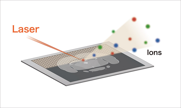
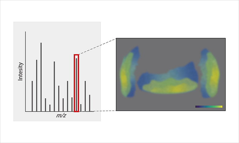
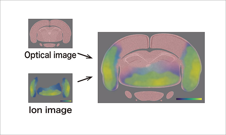
The MSI function is available as an option for mass spectrometers, and the results obtained depend on the instrument specifications. For instance, if an instrument has high mass resolution, the MSI data can also be acquired with similarly high mass resolution. In addition, MSI typically provides ion images (molecular distributions) at a spatial resolution ranging from a few to several tens of micrometers. While the measurement parameters that contribute to resolution vary depending on the method, in the case of laser desorption/ionization, the laser irradiation diameter and raster pitch are adjusted according to the purpose.
Features and challenges of mass spectrometry imaging
Features
- Intuitive understanding through images
Ion images provide information in an easily visualized form. Although it is challenging for non-experts to interpret mass spectra, ion images can facilitate understanding. Large-area imaging such as at the scale of a microscope slide allows for intuitive grasp of component distribution at a scale close to that of the naked eye. - Simultaneous imaging of multiple components
In principle, MSI measurements do not require the use of labeling reagents to detect specific molecules. Furthermore, as described above, since mass spectral data is stored for each pixel, imaging of multiple components can be achieved in a single measurement. This is useful for evaluating the spatial correlation between a specific component and other components. - High versatility
By utilizing diverse mass spectrometry techniques, various types of imaging data can be acquired. For example, the detection of trace components and the separation of structural isomers using MS/MS or ion mobility can also be applied to MSI. Additionally, the ion images obtained may vary depending on the ionization method used. These features are highly effective for multimodal imaging analysis.
Challenges
- High initial cost
In addition to a mass spectrometer, MSI requires sample preparation equipment (such as a cryostat), which tends to result in higher initial costs than other imaging techniques. This is also one of the barriers to the wider adoption of MSI. - Trade-off between resolution and sensitivity
As the pixel size decreases, the number of ions also tends to decrease, which leads to a decline in sensitivity. When resolution is set to prioritize sensitivity (typically around 50 μm), it may become difficult to compare the results with microscope or SEM images. In recent years, efforts have been made to improve this through enhancements in mass spectrometer performance and data processing. - Limitations in quantitative analysis
Due to the nature of MSI, where the sample itself is heterogeneous, quantitative analysis remains challenging. Variations in sample surface irregularities and tissue characteristics can affect signal intensity. Particular caution is required when interpreting signal intensity and comparing signal intensity between samples. In recent years, measurement protocols and analytical approaches that take such variability into account have been proposed.
Key technologies used in MSI
MALDI
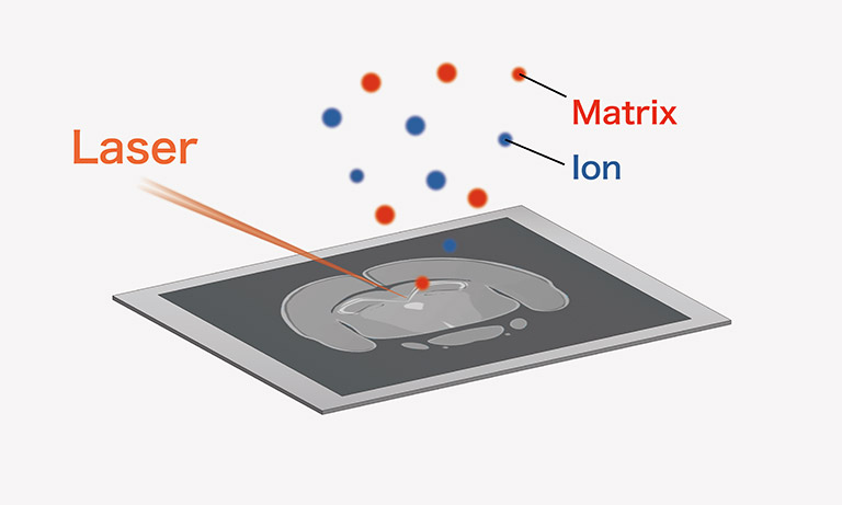
Matrix-Assisted Laser Desorption Ionization (MALDI) is currently the most widely used ionization method in MSI. A matrix, a reagent, which assists ionization, is applied to the surface of a thin section of a sample, and a UV laser is locally irradiated. The matrix absorbs the laser and facilitates the desorption and ionization of the sample, enabling ionization without significant fragmentation. MALDI is commonly combined with a time-of-flight (TOF) mass spectrometer or a quadrupole time-of-flight (QTOF) mass spectrometer.
DESI
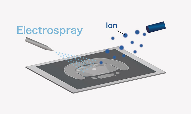
Desorption Electrospray Ionization (DESI) is a technique based on electrospray ionization to extract, desorb, and ionize sample components by spraying charged droplets. It differs from MALDI in many fundamental aspects and is rapidly gaining popularity as an alternative MSI modality . While it is commonly combined with QTOF, high-sensitivity target imaging using a triple-quadrupole (TQ) mass spectrometer has also been implemented recently.
TOF-SIMS
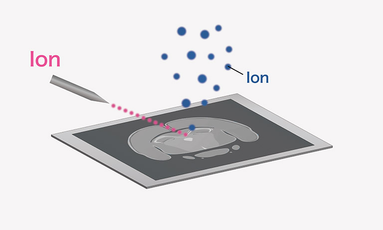
Time-of-flight secondary ion mass spectrometry (TOF-SIMS) generates mass spectra by irradiating a sample surface with primary ions and introducing the sputtered secondary ions into a TOF-MS. It is characterized by submicron-level spatial resolution and high surface sensitivity and is a widely used technique in the field of surface analysis.
Although it tends to produce more ion fragmentation than MALDI and DESI, cluster ion sources that are capable of soft ionization have been developed in recent years.
LA-ICP-MS
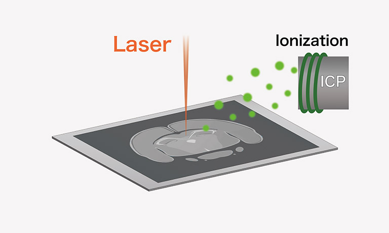
Laser ablation inductively coupled plasma mass spectrometry (LA-ICP-MS) irradiates the sample surface with a laser beam, and introduces the ablated sample into an ICP-MS. As the sample is decomposed into the atomic state within the ICP-MS, images of each element can be obtained. In biosciences, it is used for the analysis of trace metal elements in biological samples.
Sample preparation required for MSI
Thin sectioning
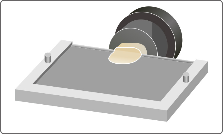
One of the sample preparations required for MSI is thin sectioning. This refers to the process of slicing a frozen sample into sections approximately 5 μm to 20 μm thick using a cryostat. It requires advanced technical skill as it is necessary to produce uniform thin sections without causing delocalization of sample components.
Examples of challenges in thin sectioning
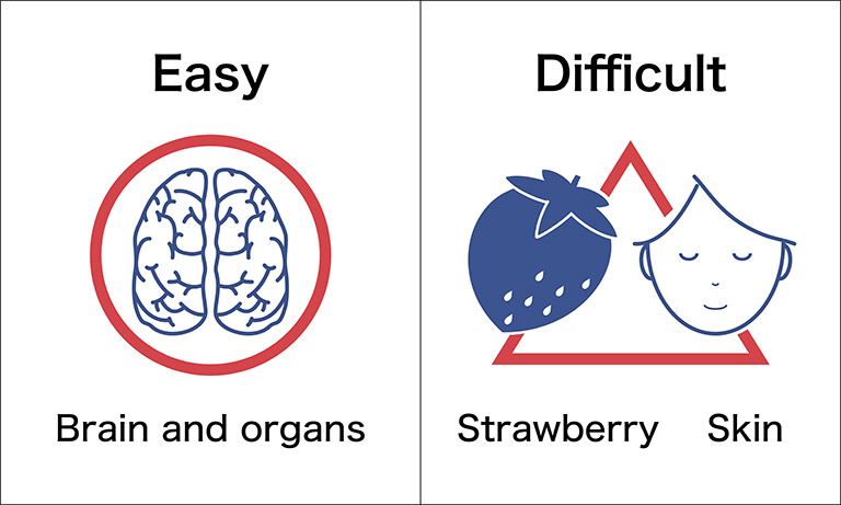
Depending on the sample, thin sectioning can be difficult due to properties such as fragility, hardness, or thinness. As measurements cannot be performed without thin sectioning, this process limits the applicability of MSI. However, the application of MSI is gradually expanding by using alternative sampling methods (such as transfer) that can replace thin sectioning.
Matrix application
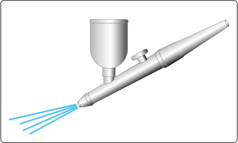
When MSI is performed using MALDI, matrix application is required in addition to thin sectioning. This process also requires specialized knowledge and experience, such as selecting a matrix compound appropriate for the sample and uniformly applying it onto the sample surface. Dedicated matrix application equipment is also available to make this easier.
Our efforts to expand the range of MSI applications
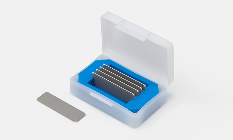
Poropare, developed by Hamamatsu Photonics, is a sample preparation support product for mass spectrometry imaging featuring a robust porous structure. By utilizing a technique called transfer sampling, which leverages the features of this product, MSI can be performed without the conventionally required thin sectioning process. This allows for MSI analysis to be applied to a wider range of samples, including those that were previously excluded from the application of MSI due to the difficulty of thin sectioning.
In addition, Poropare for MALDI is pre-coated with an ionization-assisting layer on its plate surface, enabling matrix-free analysis. This significantly simplifies sample preparation, lowers the barrier to performing MSI, and eliminates matrix-derived background noise, contributing to improvement in signal-to-noise (S/N) ratio, especially in the analysis of low-molecular-weight compounds. On the other hand, for the analysis of components that are difficult to detect under matrix-free conditions, Poropare can also be used in combination with a matrix.
*Poropare is a registered trademark of Hamamatsu Photonics K.K.
Related application

Component analysis of crops plays an extremely important role in the research and development of crop varieties as well as pesticides and fertilizers. Elucidating the chemical components contained in crops and their physiological functions is expected to promote research and development in areas such as variety improvement, the development of pesticides and fertilizers and the optimization of application conditions, thereby contributing to the improvement of crop productivity and quality. A wide range of analytical methods are used for this purpose, including spectroscopic methods and mass spectrometry. In addition to the development of analytical instruments, sample preparation processes such as extraction and separation have become increasingly sophisticated over the years, enabling the highly sensitive detection and quantitative analysis of trace components.
Related information
Mass spectrometry is a method for identifying and quantifying substances by converting them into fine ions at the atomic or molecular level through various ionization methods and then measuring their mass-to-charge ratios and quantity. Compared with other analytical methods, it provides a larger amount of information in a single analysis, making it applicable across a wide range of fields, including proteomics.
Hamamatsu Photonics offers a wide range of devices that form the core of mass spectrometers, including ion detection devices.
- Confirmation
-
It looks like you're in the . If this is not your location, please select the correct region or country below.
You're headed to Hamamatsu Photonics website for GB (English). If you want to view an other country's site, the optimized information will be provided by selecting options below.
In order to use this website comfortably, we use cookies. For cookie details please see our cookie policy.
- Cookie Policy
-
This website or its third-party tools use cookies, which are necessary to its functioning and required to achieve the purposes illustrated in this cookie policy. By closing the cookie warning banner, scrolling the page, clicking a link or continuing to browse otherwise, you agree to the use of cookies.
Hamamatsu uses cookies in order to enhance your experience on our website and ensure that our website functions.
You can visit this page at any time to learn more about cookies, get the most up to date information on how we use cookies and manage your cookie settings. We will not use cookies for any purpose other than the ones stated, but please note that we reserve the right to update our cookies.
1. What are cookies?
For modern websites to work according to visitor’s expectations, they need to collect certain basic information about visitors. To do this, a site will create small text files which are placed on visitor’s devices (computer or mobile) - these files are known as cookies when you access a website. Cookies are used in order to make websites function and work efficiently. Cookies are uniquely assigned to each visitor and can only be read by a web server in the domain that issued the cookie to the visitor. Cookies cannot be used to run programs or deliver viruses to a visitor’s device.
Cookies do various jobs which make the visitor’s experience of the internet much smoother and more interactive. For instance, cookies are used to remember the visitor’s preferences on sites they visit often, to remember language preference and to help navigate between pages more efficiently. Much, though not all, of the data collected is anonymous, though some of it is designed to detect browsing patterns and approximate geographical location to improve the visitor experience.
Certain type of cookies may require the data subject’s consent before storing them on the computer.
2. What are the different types of cookies?
This website uses two types of cookies:
- First party cookies. For our website, the first party cookies are controlled and maintained by Hamamatsu. No other parties have access to these cookies.
- Third party cookies. These cookies are implemented by organizations outside Hamamatsu. We do not have access to the data in these cookies, but we use these cookies to improve the overall website experience.
3. How do we use cookies?
This website uses cookies for following purposes:
- Certain cookies are necessary for our website to function. These are strictly necessary cookies and are required to enable website access, support navigation or provide relevant content. These cookies direct you to the correct region or country, and support security and ecommerce. Strictly necessary cookies also enforce your privacy preferences. Without these strictly necessary cookies, much of our website will not function.
- Analytics cookies are used to track website usage. This data enables us to improve our website usability, performance and website administration. In our analytics cookies, we do not store any personal identifying information.
- Functionality cookies. These are used to recognize you when you return to our website. This enables us to personalize our content for you, greet you by name and remember your preferences (for example, your choice of language or region).
- These cookies record your visit to our website, the pages you have visited and the links you have followed. We will use this information to make our website and the advertising displayed on it more relevant to your interests. We may also share this information with third parties for this purpose.
Cookies help us help you. Through the use of cookies, we learn what is important to our visitors and we develop and enhance website content and functionality to support your experience. Much of our website can be accessed if cookies are disabled, however certain website functions may not work. And, we believe your current and future visits will be enhanced if cookies are enabled.
4. Which cookies do we use?
There are two ways to manage cookie preferences.
- You can set your cookie preferences on your device or in your browser.
- You can set your cookie preferences at the website level.
If you don’t want to receive cookies, you can modify your browser so that it notifies you when cookies are sent to it or you can refuse cookies altogether. You can also delete cookies that have already been set.
If you wish to restrict or block web browser cookies which are set on your device then you can do this through your browser settings; the Help function within your browser should tell you how. Alternatively, you may wish to visit www.aboutcookies.org, which contains comprehensive information on how to do this on a wide variety of desktop browsers.
5. What are Internet tags and how do we use them with cookies?
Occasionally, we may use internet tags (also known as action tags, single-pixel GIFs, clear GIFs, invisible GIFs and 1-by-1 GIFs) at this site and may deploy these tags/cookies through a third-party advertising partner or a web analytical service partner which may be located and store the respective information (including your IP-address) in a foreign country. These tags/cookies are placed on both online advertisements that bring users to this site and on different pages of this site. We use this technology to measure the visitors' responses to our sites and the effectiveness of our advertising campaigns (including how many times a page is opened and which information is consulted) as well as to evaluate your use of this website. The third-party partner or the web analytical service partner may be able to collect data about visitors to our and other sites because of these internet tags/cookies, may compose reports regarding the website’s activity for us and may provide further services which are related to the use of the website and the internet. They may provide such information to other parties if there is a legal requirement that they do so, or if they hire the other parties to process information on their behalf.
If you would like more information about web tags and cookies associated with on-line advertising or to opt-out of third-party collection of this information, please visit the Network Advertising Initiative website http://www.networkadvertising.org.
6. Analytics and Advertisement Cookies
We use third-party cookies (such as Google Analytics) to track visitors on our website, to get reports about how visitors use the website and to inform, optimize and serve ads based on someone's past visits to our website.
You may opt-out of Google Analytics cookies by the websites provided by Google:
https://tools.google.com/dlpage/gaoptout?hl=en
As provided in this Privacy Policy (Article 5), you can learn more about opt-out cookies by the website provided by Network Advertising Initiative:
http://www.networkadvertising.org
We inform you that in such case you will not be able to wholly use all functions of our website.
Close