![]()
Products


We are actively taking measures to improve product quality levels.
Applications


Why Hamamatsu?
Resources


Support


Our company


Investors


United Kingdom (EN)
Select your region or country.
3D live cell imaging analysis of epidermal keratinocytes
Published on February 13, 2025
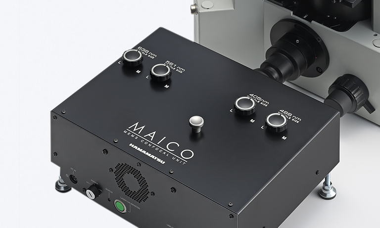
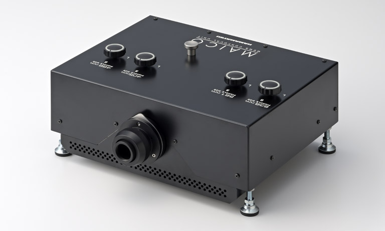
The Laboratory for Evolutionary Cell Biology of Skin, Cosmetics Course, School of Bioscience and Biotechnology, Tokyo University of Technology, is researching epidermal barrier formation mechanisms. To elucidate the mechanisms of epidermal barrier formation, it is necessary to image the epidermis in three dimensions. For this purpose, they have introduced our MAICO® MEMS confocal Unit.
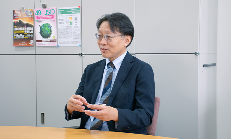
We interviewed Professor. Takeshi Matsui from the same laboratory about the background of introducing the MAICO MEMS confocal unit, his impressions of its use, and the prospects for future research.
Current research
Could you tell us about your research?
Our laboratory studies the mechanisms of adaptive evolution in the skin, particularly focusing on how the epidermal barrier is formed. The skin consists of three layers: the epidermis, dermis, and subcutaneous tissue. The epidermis is a stratified squamous epithelium made up of multiple layers, with its outermost layer being the stratum corneum (cornified layer) composed of dead cells. This stratum corneum evolved when ancient amphibians made the transition to land, and it serves as a crucial barrier that enables terrestrial vertebrates to survive on land. The stratum corneum is formed through a specialized type of cell death called corneoptosis, which occurs in the SG1 cells located in the uppermost part of the granular layer. Our laboratory has developed a live-cell imaging system using optical microscopy to observe and understand the process of corneoptosis.

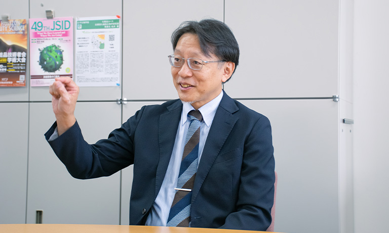
Professor Takeshi Matsui
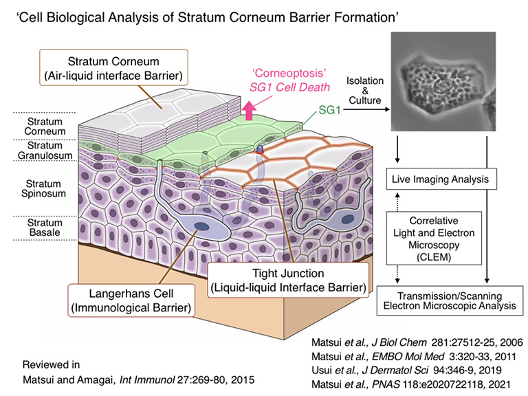
Cross-sectional view of skin epidermal tissue
Challenges in 3D live-cell imaging of the epidermis
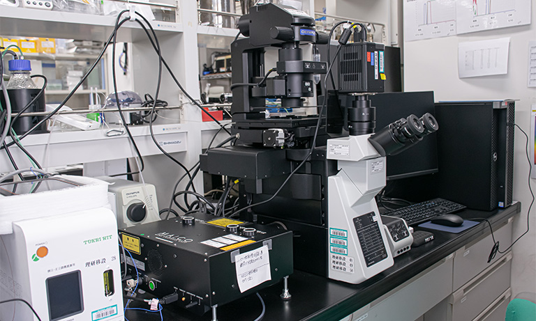
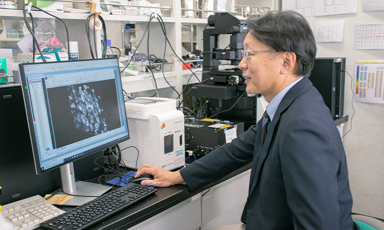
Live-cell imaging system using MAICO
Professor Matsui also uses our ORCA®-Fusion BT for regular epifluorescence imaging.
What are the challenges of live-cell imaging of the epidermis?
Since the epidermis we studied has a multilayered structure, we needed to image its structure in three dimensions. Regular fluorescence microscopes and cameras couldn't capture this 3D structure clearly, so we had to use confocal microscopy. However, confocal microscopes are expensive, making it difficult for our laboratory to purchase one. Previously, we had to rely on shared equipment available at our research institute and university.
Our experiments required time-lapse imaging to observe the process of corneoptosis in granular layer cells, sometimes necessitating continuous imaging over several days. However, the shared equipment operated on a reservation system with limited time slots. Additionally, each imaging session required considerable setup time – we needed an incubator for cell culture and had to configure settings for multi-point time-lapse imaging.
Given these circumstances, we wanted to install our confocal microscope in our laboratory so we could conduct experiments without time constraints or the hassle of repeated setup procedures.
Decisive factor for introducing the MAICO MEMS confocal unit
What made you decide to introduce MAICO?
We had been using Hamamatsu Photonics' sCMOS cameras for fluorescence imaging. While we were looking for a confocal microscope, a sales representative informed us about the newly released MAICO MEMS confocal unit, which could transform existing microscopes into confocal microscopes simply by adding it on. We first requested a demonstration.
Although I was initially concerned about the image quality and sensitivity due to its very reasonable price, I remember being surprised during the demonstration - it could capture high-quality images comparable to other companies' confocal microscopes. We ultimately decided to implement it based on several factors: its reasonable price, image quality comparable to other confocal microscopes, the ability to start with just one wavelength and add more later through its modular subunit structure, and the compact size of the equipment.
Another deciding factor was MAICO's unique feature of simultaneous multi-wavelength observation without crosstalk. Since our laboratory conducts live cell imaging, we wanted to capture multiple wavelengths simultaneously. Usually, this causes wavelength bleeding problems, but MAICO solved this issue, making it extremely valuable for our work.
Recently, we have completed the installation of an XY stage for multi-point time-lapse imaging and a Z-drift compensator to maintain focal position, in addition to the MAICO MEMS confocal unit. Now that we finally have a complete setup for confocal time-lapse imaging, we're about to begin actual imaging.
Imaging examples
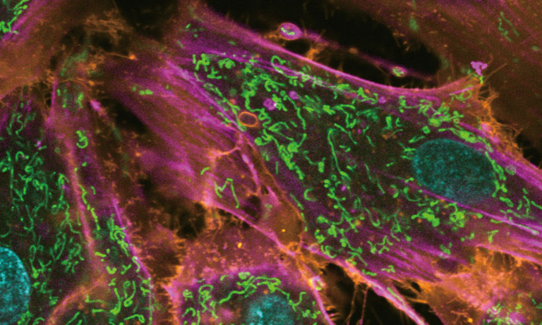
Z-stack image of mouse skin epidermal granular layer cells
After isolation of granular layer cell sheets from EGFP-expressing mouse skin, they were stained using Hoechst 33342 and Z-stack images were acquired.
Data provided by: Prof. Takeshi Matsui, Laboratory for Evolutionary Cell Biology of the Skin, School of Bioscience and Biotechnology, Tokyo University of Technology
Usability of the MAICO MEMS confocal unit
How would you feel about the usability of MAICO?
I find it very user-friendly with a simple operation. For example, while the pinhole settings don't allow for detailed adjustments, they can be set in three levels - S, M, and L - which is simple and easy to understand. This makes it smooth to explain to new students joining the laboratory each year. Additionally, for optimal imaging conditions with each lens, Hamamatsu Photonics provides a resolution simulator, which I find helpful as it allows us to adjust the Z-slice thickness according to the sample structure and thickness while referring to the simulation.
Prospects for research
Could you tell us about future research prospects?
Currently, we primarily conduct experiments using human and amphibian epidermis samples. In the future, we hope to observe and study the epidermis of amphibians and reptiles to investigate species-specific differences and evolutionary mechanisms. We plan to conduct more detailed research by combining confocal microscopy images with electron microscopy analysis and genetic information.
Since our laboratory is in the community of dermatological research, we sometimes collaborate with dermatologists and cosmetics manufacturers on various research projects, including studies on atopic dermatitis (where the skin barrier is compromised) and the evaluation of cosmetic ingredients. The live cell imaging system I'm currently developing to elucidate epidermal barrier formation mechanisms could potentially be applied to these studies as well. Therefore, we're also focusing on developing new evaluation systems.
Researcher profile

Takeshi Matsui
Professor, Laboratory for Evolutionary Cell Biology of Skin, Cosmetics Course, School of Bioscience and Biotechnology, Tokyo University of Technology
Mar. 2000
Ph. D., Graduate School of Medicine and Faculty of Medicine, Kyoto University
Apr. 2000
Group Leader, Research Group of Dermatology, KAN Research Institute, Inc.
Oct. 2006
Industry-Academia-Government collaboration Researcher, Kyoto University
Nov. 2006
Project Researcher, Graduate School of Medicine, The University of Osaka
Apr. 2007
Assistant Professor, Laboratory of Biological Science, Graduate School of Frontier Biosciences, The University of Osaka
Nov. 2007
Lecturer, Medical Top Track (MTT) Program, Medical Research Institute, Tokyo Medical and Dental University
Apr. 2011
Assistant Professor, Institute for Integrated Cell-Material Sciences (iCeMS), Kyoto University
Apr. 2013
Senior Researcher, Laboratory for Skin Homeostasis, RIKEN Center for Integrative Medical Sciences
Apr. 2016
Deputy Team Leader, Laboratory for Skin Homeostasis, RIKEN Center for Integrative Medical Sciences
Apr. 2021
Professor, Laboratory for Evolutionary Cell Biology of Skin, Cosmetics Course, School of Bioscience and Biotechnology, Tokyo University of Technology
*The content presented on this page is based on an interview conducted in October 2024.
Other case studies
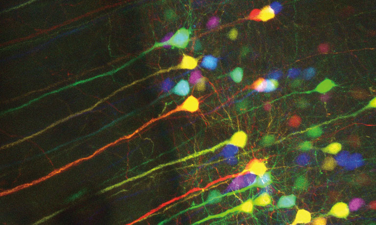
The Nano-Integration Devices and Systems Laboratory at the Research Institute of Electrical Communication (RIEC), Tohoku University, specializes in brain-inspired non-von Neumann computing and the foundational technologies for related hardware research. Within this laboratory, Associate Professor Hideaki Yamamoto's research group, the Nano-Integration Neurocomputing Systems Group, combines semiconductor microfabrication, nerve cell culture, and mathematical modeling to develop new in vitro systems for bottom-up analysis of brain functions. In these in vitro systems, cultured neurons occasionally aggregate to form a 3D structure. The group introduced the MAICO® MEMS confocal unit for 3D imaging of these aggregated neurons.


MAICO enables imaging with reduced bleed-through between wavelengths, which is an issue in multi-wavelength simultaneous observation. We will introduce how we have achieved a reduction of bleed-through.
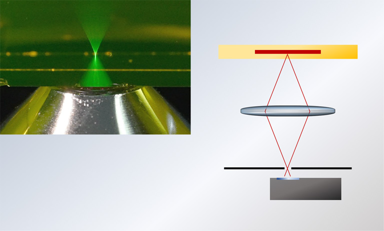
Explanation of the principles of a confocal microscope, which enables you to acquire an image that is less blurry, higher contrast, and higher resolution.

- Confirmation
-
It looks like you're in the . If this is not your location, please select the correct region or country below.
You're headed to Hamamatsu Photonics website for GB (English). If you want to view an other country's site, the optimized information will be provided by selecting options below.
In order to use this website comfortably, we use cookies. For cookie details please see our cookie policy.
- Cookie Policy
-
This website or its third-party tools use cookies, which are necessary to its functioning and required to achieve the purposes illustrated in this cookie policy. By closing the cookie warning banner, scrolling the page, clicking a link or continuing to browse otherwise, you agree to the use of cookies.
Hamamatsu uses cookies in order to enhance your experience on our website and ensure that our website functions.
You can visit this page at any time to learn more about cookies, get the most up to date information on how we use cookies and manage your cookie settings. We will not use cookies for any purpose other than the ones stated, but please note that we reserve the right to update our cookies.
1. What are cookies?
For modern websites to work according to visitor’s expectations, they need to collect certain basic information about visitors. To do this, a site will create small text files which are placed on visitor’s devices (computer or mobile) - these files are known as cookies when you access a website. Cookies are used in order to make websites function and work efficiently. Cookies are uniquely assigned to each visitor and can only be read by a web server in the domain that issued the cookie to the visitor. Cookies cannot be used to run programs or deliver viruses to a visitor’s device.
Cookies do various jobs which make the visitor’s experience of the internet much smoother and more interactive. For instance, cookies are used to remember the visitor’s preferences on sites they visit often, to remember language preference and to help navigate between pages more efficiently. Much, though not all, of the data collected is anonymous, though some of it is designed to detect browsing patterns and approximate geographical location to improve the visitor experience.
Certain type of cookies may require the data subject’s consent before storing them on the computer.
2. What are the different types of cookies?
This website uses two types of cookies:
- First party cookies. For our website, the first party cookies are controlled and maintained by Hamamatsu. No other parties have access to these cookies.
- Third party cookies. These cookies are implemented by organizations outside Hamamatsu. We do not have access to the data in these cookies, but we use these cookies to improve the overall website experience.
3. How do we use cookies?
This website uses cookies for following purposes:
- Certain cookies are necessary for our website to function. These are strictly necessary cookies and are required to enable website access, support navigation or provide relevant content. These cookies direct you to the correct region or country, and support security and ecommerce. Strictly necessary cookies also enforce your privacy preferences. Without these strictly necessary cookies, much of our website will not function.
- Analytics cookies are used to track website usage. This data enables us to improve our website usability, performance and website administration. In our analytics cookies, we do not store any personal identifying information.
- Functionality cookies. These are used to recognize you when you return to our website. This enables us to personalize our content for you, greet you by name and remember your preferences (for example, your choice of language or region).
- These cookies record your visit to our website, the pages you have visited and the links you have followed. We will use this information to make our website and the advertising displayed on it more relevant to your interests. We may also share this information with third parties for this purpose.
Cookies help us help you. Through the use of cookies, we learn what is important to our visitors and we develop and enhance website content and functionality to support your experience. Much of our website can be accessed if cookies are disabled, however certain website functions may not work. And, we believe your current and future visits will be enhanced if cookies are enabled.
4. Which cookies do we use?
There are two ways to manage cookie preferences.
- You can set your cookie preferences on your device or in your browser.
- You can set your cookie preferences at the website level.
If you don’t want to receive cookies, you can modify your browser so that it notifies you when cookies are sent to it or you can refuse cookies altogether. You can also delete cookies that have already been set.
If you wish to restrict or block web browser cookies which are set on your device then you can do this through your browser settings; the Help function within your browser should tell you how. Alternatively, you may wish to visit www.aboutcookies.org, which contains comprehensive information on how to do this on a wide variety of desktop browsers.
5. What are Internet tags and how do we use them with cookies?
Occasionally, we may use internet tags (also known as action tags, single-pixel GIFs, clear GIFs, invisible GIFs and 1-by-1 GIFs) at this site and may deploy these tags/cookies through a third-party advertising partner or a web analytical service partner which may be located and store the respective information (including your IP-address) in a foreign country. These tags/cookies are placed on both online advertisements that bring users to this site and on different pages of this site. We use this technology to measure the visitors' responses to our sites and the effectiveness of our advertising campaigns (including how many times a page is opened and which information is consulted) as well as to evaluate your use of this website. The third-party partner or the web analytical service partner may be able to collect data about visitors to our and other sites because of these internet tags/cookies, may compose reports regarding the website’s activity for us and may provide further services which are related to the use of the website and the internet. They may provide such information to other parties if there is a legal requirement that they do so, or if they hire the other parties to process information on their behalf.
If you would like more information about web tags and cookies associated with on-line advertising or to opt-out of third-party collection of this information, please visit the Network Advertising Initiative website http://www.networkadvertising.org.
6. Analytics and Advertisement Cookies
We use third-party cookies (such as Google Analytics) to track visitors on our website, to get reports about how visitors use the website and to inform, optimize and serve ads based on someone's past visits to our website.
You may opt-out of Google Analytics cookies by the websites provided by Google:
https://tools.google.com/dlpage/gaoptout?hl=en
As provided in this Privacy Policy (Article 5), you can learn more about opt-out cookies by the website provided by Network Advertising Initiative:
http://www.networkadvertising.org
We inform you that in such case you will not be able to wholly use all functions of our website.
Close